EMS MEd Blog

Can Situational Awareness Be Taught?

Article by Karlee De Monnin

CASE

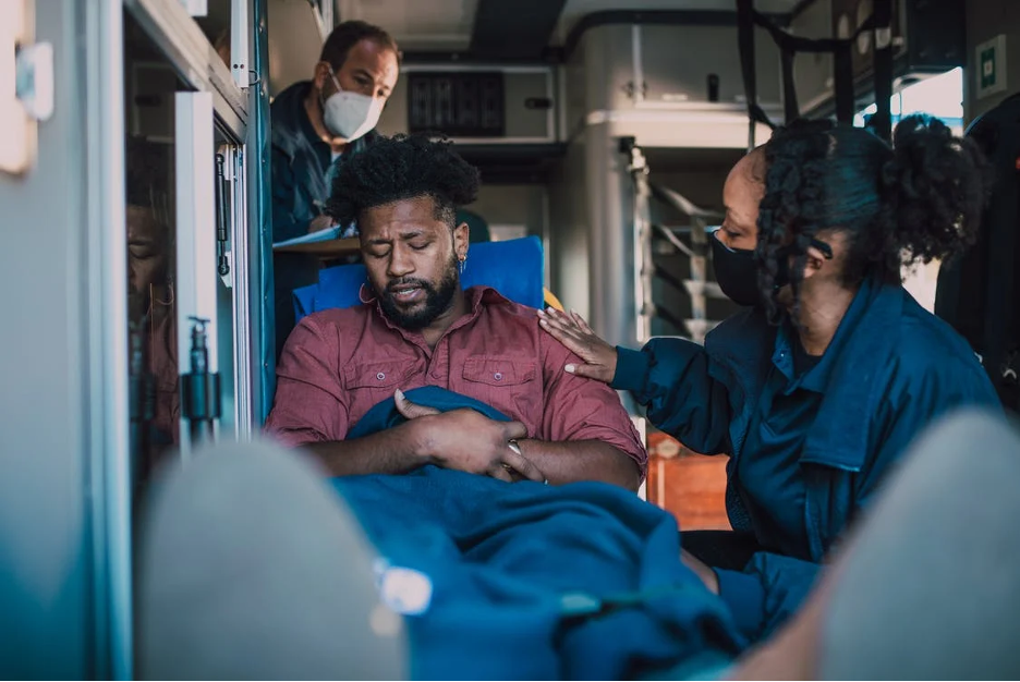
During my first ambulance ride-along as a fourth-year medical student on an EMS elective, my unit responded to a call for abdominal pain. On arrival to the apartment building, we stepped inside to find a tearful, but otherwise well-appearing young woman lying on the floor of her living room.

She was an otherwise healthy 23-year-old who called us for abdominal pain and vomiting. She noted a positive home pregnancy test several days prior, for which she hadn’t yet sought prenatal care. Her abdominal pain, localized to the left lower quadrant, had been present for several days, but she had become severely nauseated and vomited several times over the past hour since awakening that morning. The paramedics assisted the patient into the ambulance. Her vitals were stable, and the team placed an IV. We began transport to the hospital.

With the truck in motion, the paramedic focused his attention on his computer screen, asking the patient several questions and inputting her information. All the while, sweat began to bead up along the patient’s hairline. Glancing up, the paramedic commented on her increasing clamminess and turned on the air conditioning before turning back to his computer.

I was focused on exploring the truck as it was my first time transporting a patient in an ambulance. As seconds passed, the patient began squirming in her seat and moaning. This quickly progressed to dry heaving. At this point, the paramedic jumped up from his seat, lunged over the patient to slide open a cabinet above my head and obtained an emesis bag. The bag made it just in time. “We’re out of Zofran, unfortunately,” he murmured to me.

REFLECTION

At first glance, this might appear to have been a relatively smooth encounter. We picked up a patient, evaluated her, and transported her safely to the hospital. And yet, there’s a tiny detail in this case that highlights a potential area for improvement: this patient had established a pattern of vomiting prior to our arrival on scene, of which we were fully aware. In the rig, she started showing clear signs that she was about to have another episode of emesis, and yet, we failed to get her an emesis receptacle until the very last second, just before it was too late. We lacked appropriate situational awareness, and narrowly avoided the consequences.

Situational awareness, of which anticipation is a major component, relies to a large degree on experience. This is a skill that is built upon time and mistakes – the team in the back of the truck that day was comprised of a relatively newer paramedic, and me, a medical student riding along in an ambulance for the first time. I was in a completely new environment and experienced the consequences of an elevated cognitive burden. Neither of us anticipated emesis until it was almost too late.

To a team of more experienced prehospital clinicians, it may have been second nature to reach for an emesis bag upon entry to the vehicle. There is no substitute for experience. Yet, regardless of experience level, it is important to consider how EMS education can teach situational awareness. This may be critically important in complicated clinical scenarios, such as being able to predict the trajectory of an unstable patient and intervene appropriately, or being able to recognize a lack of scene safety and take appropriate precautions before an accident happens. The skill of anticipation is broadly applicable across prehospital care, and is likely to be amplified in stressful situations, during which a person’s cognitive load is increased.

Since it is impossible to staff all response vehicles with only the most experienced of providers, how can we best prepare newer prehospital clinicians?

LITERATURE REVIEW

Does research show situational awareness can be taught or can it only be gained through experience? In the hospital, situational awareness is an important factor in human error analysis, a critical aspect of healthcare, given that medical errors are a remarkably common cause of death. There is some literature on situational awareness across clinical settings, and a small number of studies in the prehospital setting, which can be loosely extrapolated to offer insight into effective EMS training.

From a research perspective, the concept of situational awareness was initially developed in the aviation sector, but has since been applied broadly to other systems, including healthcare (Williams 2013). Justin Hunter and colleagues (2019) describe situational awareness as “being aware of what is happening around you, understanding what that information means now, and predicting what it will mean in the future.” In the example above, situational awareness might then be explained as recognizing the initial chief complaint of vomiting and abdominal pain, along with the patient’s increasing diaphoresis and discomfort en route.

Expanding on their definition above, Hunter and colleagues (2019) offer several theoretical frameworks by which situational awareness can be understood, of which they argue that Mica Endsley’s three-level framework of situational awareness (comprised of identification, interpretation, and prediction) is most applicable to paramedicine. Because situational awareness is a form of cognitive processing, it is unsurprisingly difficult to quantify and study. Hunter and colleagues have attempted to put forth additional studies on the topic of situational awareness, using Endsley’s framework as a guide. For example, they conducted a pilot study (2021) which considered the situational awareness of paramedic students during a simulation.  Situational awareness was low and a common theme identified was the lack of an organized approach to the situation. Inability to achieve “prediction” level situational awareness was limited also by stress and failure of recognition/perception.

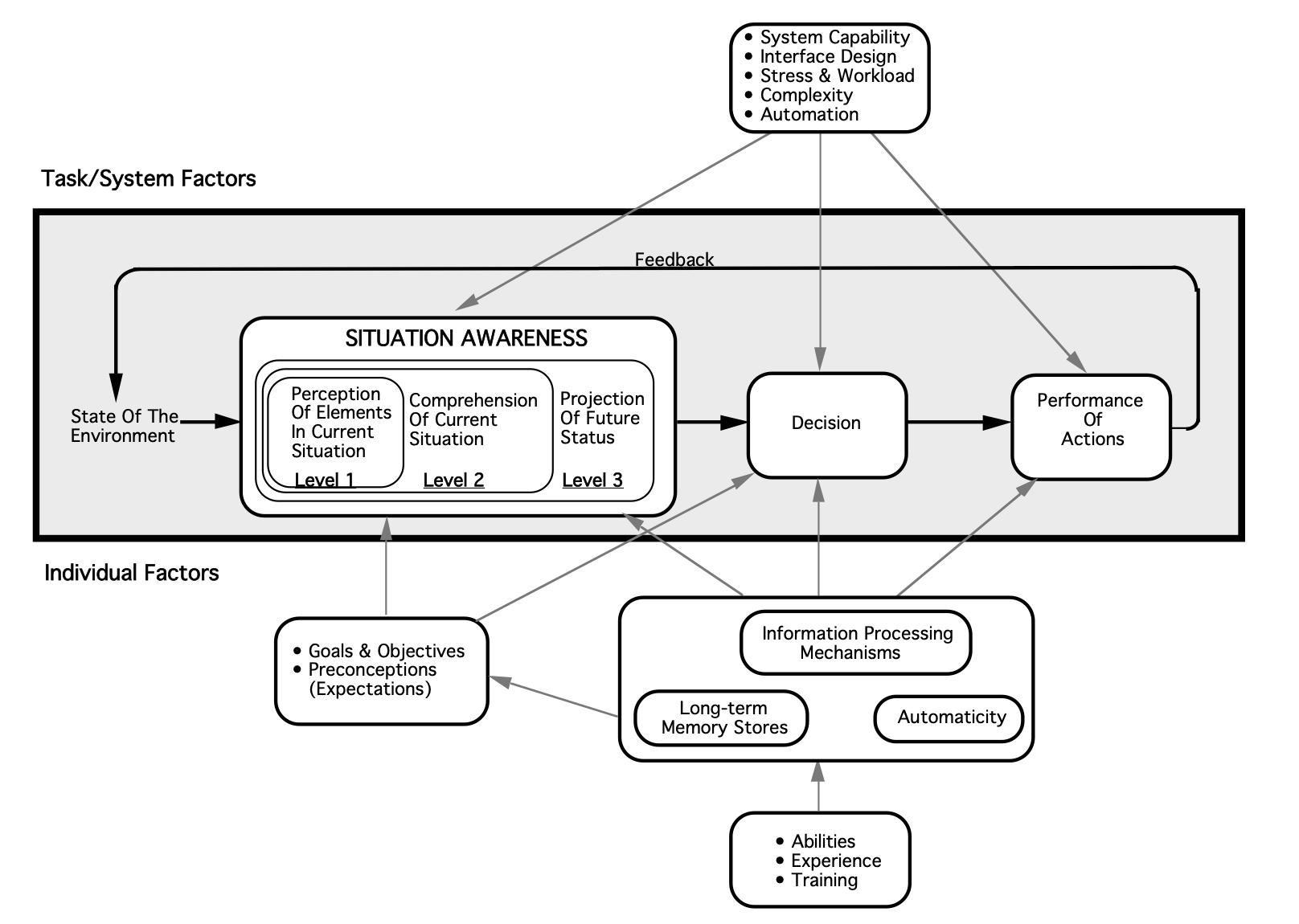
Image from Endsley, M. R. (1995). Toward a theory of situation awareness in dynamic systems. Human factors37(1), 32-64.

A small number of validated techniques have been employed for measuring situational awareness in healthcare. For example, Endsley developed the Situation Awareness Global Assessment Technique (SAGAT), which has been employed to evaluate trauma skills among medical trainees (Hogan 2006) and recognition of patient deterioration among student nurses (Kinsman 2009, Cooper 2019). This test requires halting a simulated scenario to assess participant insight into situational awareness, utilizing standardized reflective questions. An alternative metric, the Situation Present Assessment Method (SPAM), has also been employed (Williams 2013). Additionally, some interest has been expressed in examining eye tracking as a means of measuring situational awareness, though the efficacy of this, as well as its utility in clinical education, remains to be seen (Williams 2013). Little of this research has touched the prehospital arena.

Within the prehospital setting, Hunter’s group has recently started to examine educational strategies to modify situational awareness, with a statistically significant increase in situational awareness with an educational intervention in a quasi-experimental before-after study in 2022. The educational tool employed by Hunter and colleagues focused on elements of Endsley’s situational awareness model and some adapted principles from aviation, Crew Resource Management, designed to improve situational awareness.

Though the study is small, there is something to be said for acknowledging the potential value of education on situational awareness in paramedic training. The limited research on this topic is illustrative of how difficult it is to quantify and study. In the absence of clear, compelling evidence, an important question arises as to how medical directors can identify the need for targeted teaching and feedback of nontechnical skills like this, as they will not necessarily be reflected on chart review, and whether there is even value in broadly incorporating education on this skill.

CONCLUSION

Situation awareness education and training for prehospital clinicians and scenarios needs additional investigation. As research continues, we may discover evidence for interventions to improve situational awareness. Although there are time and financial costs to education and training, it important to remember that situational awareness itself is critical to prehospital patient care.

REFERENCES

  • Cooper S, Kinsman L, Buykx P, McConnell-Henry T, Endacott R, Scholes J. Managing the deteriorating patient in a simulated environment: nursing students' knowledge, skill and situation awareness. J Clin Nurs. 2010;19(15-16):2309-2318. doi:10.1111/j.1365-2702.2009.03164.x

  • Hogan MP, Pace DE, Hapgood J, Boone DC. Use of human patient simulation and the situation awareness global assessment technique in practical trauma skills assessment. J Trauma-Injury Infect Crit Care. 2006;61(5):1047–1052.

  • Hunter, J., Porter, M., & Williams, B. (2019). What Is Known About Situational Awareness in Paramedicine?: A Scoping Review. Journal of allied health48(1), e27–e34.

  • Hunter, J., Porter, M., Phillips, A., Evans-Brave, M., & Williams, B. (2021). Do paramedic students have situational awareness during high-fidelity simulation? A mixed-methods pilot study. International emergency nursing56, 100983. https://doi.org/10.1016/j.ienj.2021.100983

  • Hunter, J., Porter, M., Cody, P., & Williams, B. (2022). Can a targeted educational approach improve situational awareness in paramedicine during 911 emergency calls?. International emergency nursing63, 101174. https://doi.org/10.1016/j.ienj.2022.101174

  • Kinsman, L., Endacott, R., Cooper, S. J. R., Scholes, J., Buykx, P., & McConnell-Henry, T. E. (2009). Situational awareness of patient deterioration in a simulated environment.

  • Williams, B., Quested, A., & Cooper, S. (2013). Can eye-tracking technology improve situational awareness in paramedic clinical education?. Open access emergency medicine : OAEM5, 23–28. https://doi.org/10.2147/OAEM.S53021

About the Author:

Karlee is an emergency medicine-bound fourth-year medical student at Washington University in St. Louis. She has a strong clinical interest in medical education. Outside the hospital, she can be found running with her dog, baking, or getting lost in a book.

Editing by James Li, MD

Commercial Tourniquet Use in Pediatrics

Article by Veronica “Vee” Smith, MD

Case

It’s 11 o’clock in the morning on a sunny autumn day. Your radio alerts you about a mass casualty event and you are then dispatched to what turns out to be a school shooting. The estimated casualty count is over 20 with an unknown number of injured victims on the scene, their ages are estimated to range from 5-58 years of age. As your partner begins to drive light and sirens to the scene, several questions quickly race through your mind “Do I have enough tourniquets”, “Can I use a commercial tourniquet on a pediatric patient? And if so, is there an age cutoff?”. Before you know it you and your partner have arrived on the scene.

Introduction

Traumatic injuries are the leading cause of death among children and adolescents aged 1-17 years of age in the United States. Many of those deaths have been attributed to motor vehicle accidents, homicide, suicide and non-accidental trauma [1]. In 2020, penetrating trauma from firearm injuries became the leading cause of traumatic deaths in the pediatric population, surpassing deaths caused by motor vehicle accidents [1]. Traumatic brain injuries are the most common injury complex associated with death in children with traumatic injuries, followed by anoxia and hemorrhage [2]. Most deaths occur within the first 24 hours of the primary injury emphasizing the importance of interventions performed in the prehospital setting and the immediate hospital care in the emergency department [2]. Death due to hemorrhagic shock is more commonly associated with gunshot wounds with death occurring earlier in the first 24 hours in comparison to deaths from TBIs and anoxia [2]. As school shootings have continued to be on the rise in the US, there has been increased focus on hemorrhage control in the prehospital setting due to its significant morbidity and mortality. Data on tourniquet use in the pediatric population is currently sparse, much of the information that has been used to support the use of tourniquets in this population has been provided from research that has focused on the adult population or children injured in military war zones.

Literature Review

A study conducted at a combat support hospital in Iraq compared the outcomes of patients with hemorrhaging extremity injuries who had tourniquets applied in the prehospital setting vs tourniquet placement in the Emergency Department. Survival rates were higher in patients who received pre-hospital tourniquet placement [3]. The survival rate was also higher if the tourniquet was placed prior to the onset of shock, 90% of those patients survived their injuries in comparison to only 18% of those with tourniquets placed after the onset of shock [3]. Likewise, domestic studies involving tourniquet use in traumatic extremity injuries have shown similar results. A retrospective study at a level 1 trauma center in New Orleans examined commercial tourniquet use for penetrating extremity trauma over an 8-year period. Patients with a tourniquet were compared to those without. In comparison to the cohort without tourniquets, those who received tourniquets: required fewer blood transfusions, had higher systolic blood pressures (prehospital and on arrival to the ED), lower incidence of shock on arrival to the ED and shorter length of hospital stay [4]. In addition, the non-tourniquet cohort a higher incidence of fasciotomy and secondary amputation [4].

Many of the current commercial tourniquet styles have been used and tested in the military, with most of the use occurring in patients over the age of 18. For a tourniquet to function properly, it must be tightened to apply adequate circumferential pressure to halt blood flow distally. Some commercial tourniquets have a rigid mechanical system (i.e. windlass or ratchet) used to tighten the tourniquet onto an extremity; this can cause difficulty in fitting the tourniquet onto extremity circumferences that are smaller than the mechanism. Pediatric limb circumferences are smaller than that of an adult, which raises the question: Can enough circumferential pressure be applied to stop bleeding in small children? 

A 2019 study examined the use of the Combat Application Tourniquet (CAT) in sixty children (aged 6-16 years) with a goal in determining whether the CAT would be able to apply enough pressure to occlude extremity arterial blood flow. The CAT was applied to an upper arm and thigh while peripheral pulses were monitored by Doppler. The number of windlass turns to occlude arterial blood flow were also recorded (maximum allowed was 3 turns (1080 degrees)). The CAT was found to have a 100% success rate in occluding arterial blood flow in the upper extremities tested [5]. In comparison the success rate in the lower extremity was 93% [5]. There was an increase in the number of windlass turns required to occlude blood flow as patients age and BMI increased [5]. Though this study provided demonstration that CATs could successfully be used in pediatric human subjects, the question of whether there is an age limit for successful use in commercial tourniquets remains. A 2020 study also examined the application of CATs to pediatric upper and lower extremities, this time examining children aged 2-7 years [6]. Although the sample size was smaller (24 extremities tested), the study found a 100% success rate in occluding arterial blood flow in both upper and lower extremities [6]. The smallest arm circumference and leg circumference included in the study were 13cm and 24.5cm respectively [6]. It may be possible that commercial tourniquets may be successful in children under the age of 2, but currently there has not been human subject research performed on this age group to provide that evidence.

Conclusion

High profile mass casualty events such as the Boston Marathon bombing have increased public interest in tourniquet use. With educational campaigns such as Stop the Bleed, commercial tourniquet use in the prehospital setting will likely increase and provide more information on tourniquet use and outcomes in the pediatric population. Although commercial tourniquets such as the CAT were designed for adult use particularly that of servicemen and servicewomen, at least two studies have shown that they can be successfully applied to pediatric patients aged 2-16 years [5,6]. The patients in these studies were volunteers with no extremity trauma, prehospital research could establish more efficacy of commercial tourniquet use in pediatric patients experiencing extremity trauma with exsanguinating hemorrhage. Despite limited research data the Pediatric Trauma Society supports (PTS) the usage of tourniquets in the prehospital setting during the resuscitation of pediatric patients with severe extremity trauma and hemorrhage [7]. In addition, PTS also supports the Stop The Bleed campaign, placing emphasis on prehospital hemorrhage control by use of direct pressure, wound packing with hemostatic gauze and tourniquet use.

References

  1. Centers for Disease Control and Prevention. Web-based Injury Statistics Query and Reporting System. https://www.cdc.gov/injury/wisqars/index.html

  2. Theodorou CM, Galganski LA, Jurkovich GJ, Farmer DL, Hirose S, Stephenson JT, Trappey AF. Causes of early mortality in pediatric trauma patients. J Trauma Acute Care Surg. 2021 Mar 1;90(3):574-581. doi: 10.1097/TA.0000000000003045. PMID: 33492107; PMCID: PMC8008945.

  3. Kragh JF Jr, Littrel ML, Jones JA, Walters TJ, Baer DG, Wade CE, Holcomb JB. Battle casualty survival with emergency tourniquet use to stop limb bleeding. J Emerg Med. 2011 Dec;41(6):590-7. doi: 10.1016/j.jemermed.2009.07.022. Epub 2009 Aug 31. PMID: 19717268.

  4. Smith AA, Ochoa JE, Wong S, Beatty S, Elder J, Guidry C, McGrew P, McGinness C, Duchesne J, Schroll R. Prehospital tourniquet use in penetrating extremity trauma: Decreased blood transfusions and limb complications. J Trauma Acute Care Surg. 2019 Jan;86(1):43-51. doi: 10.1097/TA.0000000000002095. PMID: 30358768.

  5. Harcke HT, Lawrence LL, Gripp EW, Kecskemethy HH, Kruse RW, Murphy SG. Adult Tourniquet for Use in School-Age Emergencies. Pediatrics. 2019 Jun;143(6):e20183447. doi: 10.1542/peds.2018-3447. Epub 2019 May 7. PMID: 31064797.

  6. Kelly JR, Levy MJ, Reyes J, Anders J. Effectiveness of the combat application tourniquet for arterial occlusion in young children. J Trauma Acute Care Surg. 2020 May;88(5):644-647. doi: 10.1097/TA.0000000000002594. PMID: 31977996.

  7. Cunningham A, Auerbach M, Cicero M, Jafri M. Tourniquet usage in prehospital care and resuscitation of pediatric trauma patients-Pediatric Trauma Society position statement. J Trauma Acute Care Surg. 2018 Oct;85(4):665-667. doi: 10.1097/TA.0000000000001839. PMID: 29462083.

Author Bio: Dr. Veronica "Vee" Smith is a second year Pediatric Emergency Medicine Fellow at St. Louis Children's Hospital/Washington University in St. Louis. She enjoys cooking, writing, playing & watching basketball when she is not working. 

Website Editing and Layout by James Li, MD

Complexity

EMS Perspectives: An OpEd Page on the History and Future of EMS

By Clayton Kazan, MD, MS, FACEP, FAEMS

So we are about 54 years into the pilot project that is EMS and paramedicine.  That we would even exist, much less thrive, years later, was viewed by many as highly improbable at the time.  The EMS system, in its inception, was a complicated system with a lot of moving parts.  It wasn’t simple, but it was fairly linear.  Call was made to the Fire Department (or EMS, as 911 did not exist), a response was dispatched, units arrived on scene, they provided whatever assessment and treatment were appropriate (rudimentary by today’s standards), they notified the hospital, and they transported.  The system of today has moved from complicated to extremely complex, affecting each of the steps above. 

One of the notable exceptions, at least until recently, was the 911 system.  We reduced the number of phone numbers down to a single, 3 digit number.  This is a terrific example of system leadership advocating to reduce system complexity.  911 is in place throughout the US and enables you to access the appropriate local resources without knowing the local telephone numbers.  From rotary phone to iPhone, the 911 system remained simple to the user.  Now, with the advent of 988, we are reintroducing system complexity, and not just by giving the public a second number to dial.  Bridges between the 911 and 988 system need to be developed to appropriately move traffic when the public dials the wrong number.  But, I digress, because at every other step above, through system entropy and evolution, we have introduced complexity exponentially.  From emergency medical dispatch to tiered dispatch to specialty response units to sophisticated assessment tools and new treatments, to new methods of hospital notification, and to the hyperspecialization of receiving hospitals, our system has become quite a wonderful beast.  With every layer of complexity that is added, we create new ways for the systems to fail, but we have also created an infinite way for the EMS system to aid.

So, with all of that said, the question that I have been pondering and what drove me to my laptop on my day off was this…has the complexity of our operations failed to keep up with the growing complexity demanded of it by the communities we serve?  We keep being asked to do more than EMS, to the point that EMS and prehospital care do not even seem to be the appropriate terms for what we do anymore because, so often, the E does not apply, and not all care that we provide should be prior to arriving at a hospital.  Really, what we have become is mobile health, with traditional EMS existing as a service line under that umbrella.  Nothing made this clearer than the pandemic response.  EMS (or whatever we decide to call it) rose to the occasion and demonstrated amazing resilience and adaptability when so much of the healthcare system crumbled around it.  We were the innovators.  Why?  Because we are still a pilot project.  Traditional healthcare has been around for 2400 years, and it is more rigid, yet brittle.  54 years in, EMS is still trying to find and define its niche, which is why the adage, “if you’ve seen one EMS system, you’ve seen one EMS system” applies.  You can’t say that for hospitals, Emergency Departments, clinics, etc.  They’re more like Starbucks…pretty similar but not exactly identical and very expensive to visit.

So, what do I mean by this complexity gap?  The cracks exposed in our healthcare system from the pandemic are far from repaired, and it remains to be seen how traditional healthcare will morph as it recovers.  But, we have true crises of mental health, substance use, homelessness, aging population, and eroding access to all forms of scheduled medical care, and I do not use the word “crisis” haphazardly.  We have more care facilities, of all kinds, calling us because they can’t get access to other forms of healthcare and are unwilling to shoulder the liability of watchful waiting when a free solution (at least to them) exists just three digits away.  Our pace of innovation to meet these needs has been unable to keep up, and the end result has been more and more call volume driven, mostly, by low to medium acuity patients.  As we seek new solutions, we run into perpetual problems with funding, reimbursement, and outright pushback other medicine stakeholders, who lack any granular solutions, seeking to contain any perceived expansion of the mobile health mission and scope.

Complexity can definitely be problematic, but it also generates a broader repertoire of solutions.  We need to take advantage of our relative youth, amongst the rest of the house of medicine, with all of the optimism and adaptability, and keep advocating for innovative solutions, new connections to care, new treatment modalities, new delivery models, and new transport destinations.  The going will be hard, but the alternative is the unfettered crumbling of our existing healthcare system and its growing inability to meet the needs of its constituency.  We have come a long way in 54 years, but we have a long, hard road in front of us as well.

Website Layout and Graphic by EMS MEd Editor James Li, MD

Calcium in Out-of-Hospital Cardiac Arrest

Article by Erin Lincoln, MD

Case Scenario:

You are dispatched to a 68-year-old male in cardiac arrest.  His family has been performing bystander, and report that he suddenly collapsed just a few minutes ago. CPR is taken over by responding crews, and he is placed on a cardiac monitor/defibrillator. He is found to be in ventricular fibrillation (VF).  After several cycles of defibrillation, epinephrine, and amiodarone, the patient remains in cardiac arrest.  The medic on scene calls on-line medical control to ask for advice, and specifically asks if calcium can be given, as she has “seen it work before” to get pulses back as a “last ditch effort.”

Background:

Calcium chloride or gluconate was originally utilized in cardiac arrest resuscitation in the 1950’s after a single study was published in 1951 (Kay & Blalock, 1951). However, evidence emerged in the 1980’s demonstrating that calcium chloride had no effect on return of spontaneous circulation (ROSC) rates, and in fact could be detrimental (Landry, Foran, & Koyfman, 2014). Current AHA guidelines do not recommend routine use of calcium in cardiac arrest (Panchal, et al., 2020). Calcium acts as a vasopressor and inotropic agent (Lindqwister, et al., 2020) thus lending itself to a potential drug for cardiac arrest. Calcium is also frequently used in the treatment of hyperkalemia, calcium channel blocker overdose, hypermagnesemia, and hypocalcemia and may be more likely to be used when one of these diagnoses is the suspected cause of cardiac arrest. Additionally, low ionized calcium levels have been correlated with increasing mortality in sepsis and other critical illnesses in adults and children (Bora, Ramazan, Oznur, Emre, & Basar, 2021), (Sanchez, et al., 1989). Thus, calcium may be a drug considered in these and similar etiologies as appropriate due to known association with low calcium and mortality.

However, as calcium is still used for both presumed benefit in special cases, as well as a “last ditch effort” new literature continues to be published addressing the use of calcium in cardiac arrest, including a significant recent RCT. 

What does the literature say?

Since the 1980’s, literature has been routinely published regarding the use of calcium in cardiac arrest.  Several recent papers have come out, including a double blind, randomized controlled trial of calcium in cardiac arrest, and these papers are nicely summarized in a 2014 Annals of Emergency Medicine article by Landry, Foran, and Koyfman. The take home message: “Irrespective of presenting rhythm, in patients with cardiac arrest, there is no conclusive evidence that administration of calcium during cardiopulmonary resuscitation (CPR) improves survival.” This paper also notes that many of the studies were retrospective, had varied results, and that to truly answer this question, more randomized trials were needed.

Since the publication of this review, several new studies have been published including several randomized controlled trials. 

General Adult Medical Cardiac Arrest

The Calcium for Out-of-hospital Cardiac Arrest (COCA) Trial

This trial was conducted in Denmark and demonstrated through a double blind, randomized, placebo-controlled trial that calcium likely causes harm and was stopped early at a planned interim analysis due to concern for harm in the calcium arm. 397 patients were randomized- 193 received calcium, 198 received saline; of these 37 (19%) of the calcium group achieved ROSC, 53 (27%) of patients in the saline group received ROSC, risk ratio 0.72 [95% CI 0.27-1.18]. This CI does include 1; and further and further analysis of the data showed that the likelihood that calcium has a beneficial effect (e.g. risk ratio >1) was 4% for ROSC, 6% for 30 day survival, and 4% for survival with a favorable neurologic outcome at 30 days (Vallentin, et al., 2021). Visual abstract from this study:

Calcium use during cardiac arrest: A systematic review

This systematic review published in 2022 reviewed prior literature and identified that a meta-analysis was not possible due to only three available RCTs, and only one of those was considered low risk of bias. The overall conclusion was that as less than 1% of cardiac arrest etiologies fall into a group that would potentially benefit from calcium, that routine use should be avoided (Padrao, et. al., 2022).

Association between calcium administration and outcomes during adult cardiopulmonary resuscitation at the emergency department

A small retrospective study from Thailand showed again that there is no benefit to calcium given during Emergency Department resuscitation. This study also reported decreased chances of ROSC in hypocalcemic cardiac arrest patients who received calcium, and potential harm with calcium administration during traumatic arrest. This study did not account for time of administration, so survival bias may have influenced results (Wongtanasarasin, et al., 2022).

Special Cause Cardiac Arrest: Hyperkalemia and Calcium Channel Blocker Overdose

Calcium is regarded as a mainstay treatment for patients with hyperkalemia and EKG changes, and is one of two indications listed in the European Resuscitation Council guidelines on Cardiac Arrest(Lott, et al., 2021), and AHA guidelines also maintain this use (Panchal, et al., 2020). Other AHA/European guideline indications include calcium channel blocker overdose, hypermagnesemia, or hypocalcemia

The effects of calcium and sodium bicarbonate on severe hyperkalaemia during cardiopulmonary resuscitation: A retrospective cohort study of adult in-hospital cardiac arrest

This study out of Taiwan looked at known hyperkalemic cardiac arrests who received both Sodium Bicarbonate (bicarb) and calcium during in-hospital cardiac arrest. The study included 109 hyperkalemic cardiac arrest patients from 2006 through 2012. Of these, 40 (36.7%) patients achieved sustained ROSC, but only four (3.7%) patients survived to hospital discharge. Patients were grouped based on if they received: a) neither calcium nor bicarb; b) bicarb only; c) calcium only; d) calcium AND bicarb. After analysis, bicarb was positively associated with sustained ROSC when serum potassium level was <7.9 mEq/L (odds ratio [OR]: 10.51; 95% confidence interval [CI]: 1.50−112.89; p=0.03); administration of both calcium and bicarb was positively associated with sustained ROSC when serum potassium level was <9.4 mEq/L (OR: 51.11; 95% CI: 3.12−1639.16; p=0.01). This study was limited by small sample size and does NOT look at the effect of calcium alone on known hyperkalemic arrest due to the small available numbers. As no study patients survived with favorable neurologic outcome, no outcome data is available (Wang, et al., 2016).

Secondary analysis of COCA Trial

This assessed the effect of calcium in patients with PEA/ECG characteristics that could potentially have been associated with hyperkalemia and ischemia. 104 patients from the trial were found to have PEA as their last known rhythm prior to receiving the trial drug (calcium or placebo). The rhythm obtained by the defibrillator pads was analyzed for signs of hyperkalemia including loss of P waves, wide QRS complexes and large T wave amplitude.  Of these patients, 45 received calcium, 59 received placebo; 9 patients (20%) in the calcium group achieved ROSC as compared with 23 patients (39%) in the placebo group (risk ratio 0.51, 95% CI 0.26-1). While this again does not demonstrate statistical significance to imply harm, it certainly suggests that calcium may not be as helpful as previously expected when findings of hyperkalemia are present on EKG. (Vallentin, Povlsen, Granfeldt, Terkelsen, & Andersen, 2022). The forest plot for ROSC summarizes some of this data:

Pediatrics

AHA PALS guidelines recommend against routine administration of IV calcium during pediatric cardiac arrest (Topjian, et al., 2020), but IV Calcium is still used routinely in some cases in the critical care setting, such as congenital heart disease. While literature discussing prehospital administration of calcium in pediatric cardiac arrest is sparse, in-hospital literature suggests not only that calcium doesn’t demonstrate benefit, but also is associated with worse outcomes. 

Get With The Guidelines-Resuscitation (GWTG-R) Registry

This study demonstrated worse outcomes in pediatric patients with heart disease who received calcium in cardiac arrest- survival to hospital discharge was 39% in calcium recipients vs. 46% in non-recipients (P=0.02)  (Dhillon, Kleinman, Staffa, Teele, & Thiagarajan, 2022).

An editorial responding to this study does suggest that while the above paper is effective in many ways, it fails to fully account for the fact that pediatric patients who receive calcium are most likely sicker at baseline than those who do not receive calcium, and are more likely to have worse outcomes irrespective of calcium administration (Savorgnan & Acosta, 2022).

ICE-RESUScitation Project Secondary Analysis

This in-hospital analysis initially included 1,100 patients and was designed to evaluate a CPR quality improvement bundle vs usual care, researchers also found worse outcomes in patients who received calcium, INCLUDING some subgroups that had previously been hypothesized to have potential to benefit from receiving calcium during CPR including sepsis or renal insufficiency. This study attempted to mitigate bias in pre-arrest characteristics between groups by data weighting and included a PRISM (Pediatric Risk of Mortality) score when available from 2-6 hours prior to the arrest. While this study can still only prove correlation, the weighting of variables reduces bias and further supports the association of the calcium alone and the decline in outcomes (Cashen, et al., 2023). 

Take Home Points:

Calcium (chloride or gluconate) is not recommended in routine or unknown etiology cardiac arrest for both adult and pediatric patients, and this is consistent with both the AHA and European resuscitation guidelines. This continues to be supported by new literature. Special causes of cardiac arrest to include hyperkalemia and calcium channel blocker overdose, have limited data regarding efficacy but do still carry the recommendation for calcium administration.

References:

1.     Bora, C., Ramazan, K., Oznur, A. N., Emre, A. S., & Basar, C. (2021). Ionized calcium level predicts in-hospital mortality of severe sepsis patients: A retrospective cross-sectional study. Journal of Acute Disease, 10(6), 247-251.

2.     Cashen, K., Sutton, R., Reeder, R., Ahmend, T., Bell, M., Berg, R., . . . Meert, K. (2023). Calcium use during paediatric in-hospital cardiac arrest is associated with worse outcomes. In Press.

3.     Dhillon, G. S., Kleinman, M. E., Staffa, S., Teele, S., & Thiagarajan, R. (2022, November). Calcium administration during Cardiopulmonary Resuscitation for In Hospital Cardiac Arrest in Children With Heart Disease Is Associated With Worse Survival-- A Report From the American Heart Association's Get With The Guidelines- Resuscitation (GWTG-R) Re. Pediatric Critical Care Medicine, 23(11), 860-871.

4.     Kay, J., & Blalock, A. (1951). The use of calcium chloride in the treatment of cardiac arrest in patients. Surg Gynecol Obstet, 93, 97-102.

5.     Landry, A., Foran, M., & Kyofman, A. (2014, August). Does Calcium Administration During Cardiopulmonary Resuscitation Improve Survival for Patients in Cardiac Arrest? Annals of Emergency Medicine, 64(2), 187-189.

6.     Lindqwister, A. L., Lampe, J. W., Gould, J. R., Kaufman, C., Moodie, K. L., & Paradis, N. A. (2020, Sep 4). Intravenous calcium as a pressor in a swine model of hypoxic pseudo-pulseless electrical mechanical activity-a preliminary repo. Intensive Care Med Exp, 8(1), 50.

7.     Lott, C., Truhlar, A., Alfonzo, A., Barelli, A., Gonzales-Salvado, V., Hinkelbein, J., . . . Soar, J. (2021). European Resuscitation Council Guidelines 2021: Cardiac arrest in special circumstances. Resuscitation, 161, 152-219.

8.     Padrao, E., Bustos, B., Mahesh, A., Castro, M., Randhawa, R., Dipollina, C., . . . Besen, B. (2022). Calcium use during cardiac arrest: A systematic review. Resuscitation Plus, 12, 1-9.

9.     Panchal, A., Bartos, J., Cabanas, J., Donnino, M., Drennan, I., Hirsch, K., . . . Berg, K. (2020). Part 3: Adult Basic and Advanced Life Support: 2020 American Heart Association Guidelines for Cardiopulmonary Resuscitation and Emergency Cardiovascular Care. Circulation, 142, S366-S468.

10.  Sanchez, G., Venkataraman, P., Pryor, R., Parker, M., Fry, H., & Blick, K. (1989). Journal of Pediatrics, 114(6), 952.

11.  Savorgnan, F. M., & Acosta, S. P. (2022). Calcium Chloride Is Given to Sicker Patients During Cardiopulmonary Resuscitation Events. Pediatric Critical Care Medicine, 23(11), 939-940.

12.  Topjian, A., Raymond, T., Atkins, D., CHan, M., Duff, J., Jr., B. J., . . . Schexnayder, S. (2020). Part 4: Pediatric Basic and Advanced Life Support: 2020 American Heart Association Guidelines for Cardiopulmonary Resuscitation and Emergency Cardiovascular Care. Circulation, 142, S469-S523.

13.  Vallentin, M., Granfeldt, A., Meilandt, C., Povlsen, A., Sindberg, B., & Andersen, L. (2022). Effect of Calcium vs. placebo on long term outcomes in patients with out of hospital cardiac arrest. Resuscitation, 179, 21-24.

14.  Vallentin, M., Granfelt, A., C Meilandt, P. A., Sindberg, B., Holmberg, M., Iversen, B., . . . Mortensen, L. (2021, Dec 14). Effect of Intravenous or Intraosseous Calcium vs. Saline on Return of Spontaneous Circulation in Adults With Out-of-Hospital Cardiac Arrest. JAMA, 326(22), 2268-2276.

15.  Vallentin, M., Povlsen, A., Granfeldt, A., Terkelsen, C., & Andersen, L. (2022). Effect of calcium in patients with pulseless electrical activity and electrocardiographic characteristics potentially associated with hyperkalemia and ischemia- sub-study of the Calcium for Out-of-hospital Cardiac Arrest (COCA) trial. Resuscitation, 181, 150-157.

16.  Wang, C.-H., Huang, C.-H., Chang, W.-T., Tsai, M.-S., Yu, P.-H., Wu, Y.-W., . . . Chen, W.-J. (2016). The effects of calcium and sodium bicarbonate on severe hyperkalemia during cardiopulmonary resuscitation: A retrospective cohort study of adult in-hospital cardiac arrest. Resuscitation, 98, 105-111.

17.  Wongtanasarasin, W., Ungrungseesopon, N., Namsongwong, N., Chotipongkul, P., Visavakul, O., Banping, N., . . . Phinyo, P. (2022). Association between calcium administration and outcomes during adult cardiopulmonary resuscitation at the emergency department. Turkish Journal of Emergency Medicine, 22, 67-74.

Editing by Brian Miller, MD

Website Editing and Layout by EMS MEd Editor James Li, MD

Article Bites #46: Influence of Prehospital Physician Presence on Survival after Severe Trauma

Article Summary by Robert Skinner, MD

Knapp, J., Haeske, D., Boettiger, B. W., Limacher, A., Stalder, O., Schmid, A., ... & Bernhard, M. (2019). Influence of prehospital physician presence on survival after severe trauma: systematic review and meta-analysis. Journal of Trauma and Acute Care Surgery, 87(4), 978-989.

Background:

The leading cause of death in the United States for individuals under the age of 40 is trauma, according to the CDC, with an estimated death toll of greater than 160,000 people per year [1].  Because of this, attention has been placed on how to improve trauma outcomes in the prehospital setting.  One area of interest in this field is on the deployment of EMS physicians, and the practicality of their presence in the prehospital field. 

The practice of using prehospital EMS physicians is somewhat routine in many placed in Europe [2], however is still somewhat novel in the United States.  This study points out that little literature had shown a benefit of the presence of EMS physicians in the prehospital setting, despite this being a listed key priority in Emergency and Prehospital Medicine research.  This study performed a systematic literature review and meta-analysis to look at the mortality levels in severely injured patients treated by physicians in the prehospital setting compared to those treated by a paramedic led team.

Methods:

A review of the literature was performed using PubMed and Google scholar, reviewing articles published up to 2018. The authors mention that a hand search of their personal literature was performed as well.  Search criteria included studies reporting mortality or survival of severely injured patients treated either by EMS physicians or by the traditional paramedic led team.  Inclusion criteria included any study discussing patients suffering from an acute traumatic injury. Only those studies with a comparative element were included in this study.  This includes randomized control trials, matched-pairs analysis, before and after design or observation studies with a comparative element. Scores mentioned used to compare characteristics include the Injury Severity Score (ISS), Abbreviated Injury Scale (AIS), and predicted mortality. Included studies were compared using fixed effect and random effects meta-analysis using inverse variance weighting for pooling.  To account for any potential changes in care regarding ground transport vs air ambulance, a subgroup analysis was performed on studies where the effect of helicopter transport could be excluded (i.e studies with no helicopter transport in either the physician group or paramedic group or studies that had helicopter transport in both groups.)

Results:

A total of 2,249 publications were considered for inclusion in this study, and after exclusion criteria was applied, a total of 22 studies were considered eligible for the analysis.  The included studies had a pooled sample size of 54,991 patients suffering from what was considered a severe injury.  13,629 patients were treating by a team including an EMS physician, and 41,362 were treated by a traditional paramedic led team without an on scene physician. The study reports that in the overall analysis of all included studies, the odds ratios for mortality were significantly lower in the EMS physician group (OR of 0.81, 95% CI 0.71-0.92) compared to a team without EMS physician presence. 

When the analysis was subdivided to only include studies that were adjusted or matched for injury severity using one of previously mentioned scores (in other words, where injury scores between the two studies would be comparable), the OR was 0.86 (95% CI 0.73-1.01), making the result not statistically significant.  When only more recent studies (2005-2018) were included, the OR was 0.75 (95% CI 0.64-0.88) for all studies and 0.81 (95% CI 0.67-0.97) in studies adjusted for injury severity.

In the subgroup analysis of studies with comparable modes of transport (to eliminate confounder of helicopter transport), when comparing EMS physician led treatment to paramedic based, the OR for mortality was 0.80 (95% CI 0.65-1.00) in all studies and 0.74 (95% CI, 0.53–1.03) in the more recent studies.  Although not statistically significant, there is a trend towards clinical significance.

What does this study mean for EMS?

This study suggests that there may in fact be a survival benefit for severely injured trauma patients when an EMS physician is on scene and part of the treatment team. The sample size is impressive, with 22 international studies included and a patient population of greater than 54,000 patients.  Although the date of 2005 may seem somewhat arbitrary as a cutoff for “older vs newer” literature, the study does note that training in prehospital trauma management has significantly improved over the past decade, and comparing newer studies to evaluate for changes in standards of care certainly seems to strengthen this study.

Some limitations in the study include the fact that the majority of the studies included were retrospective and observational, and the fact that timing of the mortality benefit varied by study (as most studies measured mortality after 30 days or in hospital, however some measured mortality up to a year.)

EMS clinicians deliver excellent care patient care. When available; EMS physician presence seems to be beneficial as part of a team led effort for those patients who are severely injured. Additional work and research should be pursued in this topic.

References

1. Centers for Disease Control and Prevention. National Center for Health Statistics. Leding Causes of Death. https://www.cdc.gov/nchs/fastats/ leading-causes-of-death.htm. Updated March 17, 2017. Accessed March 22, 2019.

2. Wilson MH, Habig K, Wright C, Hughes A, Davies G, Imray CH. Prehospital emergency medicine. Lancet. 2015;386(10012):2526–2534.

3. Fevang E, Lockey D, Thompson J, Lossius HM, Torpo Research Collabora- tion. The top five research priorities in physician-provided pre-hospital critical care: a consensus report from a European research collaboration. Scand J Trauma Resusc Emerg Med. 2011;19:57.

Website Editing and Layout by EMS MEd Editor James Li MD

Why should quality improvement concepts be integrated into EMS education?

By Tiffany Pleasent, MD

Case Review

A 64-year-old female calls 911 for severe shortness of breath and chest pain. An ALS crew arrives on scene to find a woman who is tachypneic, hypoxic, tachycardic, and hypotensive, with diffuse rales and rhonchi throughout her lung fields. She becomes altered and severely hypoxic. The attending paramedics begin bag-valve-mask (BVM) ventilation, correct her hypoxia and hypotension, and subsequently intubate her on scene with ketamine and rocuronium. This is followed by appropriate confirmation with waveform capnography and all other appropriate adjuncts. Upon arrival to the ambulance, the EMT notices that the patient’s heart rate is 22 bpm. Shortly after they discover that she is pulseless and apneic. They begin CPR en route to the emergency department where the patient’s rhythm deteriorates into asystole. Further resuscitation is futile, and she dies.

The ER physician in the destination emergency room determines that the endotracheal tube was misplaced in the esophagus. This EMS system promotes a “just culture” that influences the crew to self-report their difficult cases to medical direction for evaluation and feedback.

The EMS medical director reviews the case and its associated monitor file which includes the timing and quality of all vital signs, including chest compression depth and rate. The monitor file shows an initial 4-phase waveform capnography of 25 mmHg for 8 breaths immediately following intubation. It further shows a subsequent loss of end-tidal waveform. This is followed by an abrupt bradycardia occurring simultaneously with rapidly dropping SpO2. The period between loss of initial waveform capnography and initiation of chest compressions was over 7 minutes. The endotracheal tube was never evaluated, removed, nor exchanged in the field.

The EMS medical director wonders if this is a “one-off” case or an indicator of more systemic issues. She decides to evaluate other similar cases and discovers that 5% of all intubations in her system lose waveform capnography and progress to cardiac arrest. She decides to implement system-level changes to manage these cases.

Keeping patient safety as our priority

Application of a systematic approach to quality improvement allows us to do the greatest good for the greatest number of people on a consistent basis. The case above describes a system with multiple unrecognized failed airways (UFAs), some likely resulting in patient harm. Fortunately, the medical director and the service leadership have a committed desire to implement change.

Several considerations arise:

  • How does the medical director develop ideas and choose what she wants to change in order to decrease UFAs?

  • How should she know what to monitor?

  • Should she use qualitative or quantitative data to drive system change?

  • How frequently should she be analyzing the data?

  • How does she appropriately analyze the data in order to obtain the right information? When should she abort a change idea?

  • How does she know that an improvement truly is an improvement?

Both EMS physician and EMS clinician involvement are required in this process, and the system is best served when both understand key quality management principles.

What are some key quality management principles that are applicable to EMS?

The Model for Improvement

The Institute of Medicine’s (now known as the National Academy of Medicine) Model for Improvement was initially adopted from the Associates in Process Improvement as a commonly used framework for healthcare providers to improve their systems [5,7,8]. It requires clinicians a) to create a measurable aim, b) to establish quantifiable measures that can be used to evaluate improvement over time, and c) to develop change ideas to implement. This is followed by small-scale tests of change using PDSA (plan-do-study-act) cycles that are scalable if the cycle is successful.

The difference between quality improvement and quality assurance

These two terms are frequently used interchangeably, albeit incorrectly. Quality assurance describes the detection of an error after the error has occurred [8]. Quality improvement describes evaluation of the system to ensure it appropriately produces the desired results and prioritizes error avoidance. Quality improvement is proactive whereas quality assurance is reactive, and both are important.

Case Review Application

The medical director decides to apply small tests of change to improve her system. She creates an aim statement, establishes quantifiable outcome measures, and develops several change ideas. Following implementation of each change idea, she says “show me the data!”

PDSA Cycles

#1 – An e-mail memo is sent to the EMS system reminding clinicians to establish and monitor continuous waveform capnography of all airway devices while en-route to the hospital and during patient movement and handoffs. After one month, there is no change in the system’s UFA outcome measures.

Model for Improvement: PDSA Cycle (Institute for Healthcare Improvement)

#2 – A podcast is developed explaining waveform capnography and the importance of continuous monitoring of advanced airways. Additionally, a protocol revision is made outlining advanced airway confirmation and management requirements.  Unfortunately, there is minimal change in the UFA outcome measures.

#3 – To identify further change theories, the medical director facilitates a case review with the EMS clinicians involved in each of the UFA cases that resulted in cardiac arrest. Nearly all the crews recall difficulty seeing the cardiac monitor in their ambulances due to the monitor mount being poorly positioned. Furthermore, they report their monitor never alarmed that capnography was lost. Clinicians provide recommendations for better placement of the mount in the ambulance and the change is made to the entire fleet. Crews report in the following weeks a significant improvement in their ability to monitor their patient.

#4 – The cardiac monitor settings are reviewed and nearly all alarms have been turned off by crews because of frequent inappropriate alarms resulting in alarm fatigue. The team involves EMS clinicians to identify the most important alarms to maintain, including the capnography apnea alarm, and an admin password is established to prevent these settings from being changed. These monitor changes are trialed by a group of crews for a few shifts and additional small refinements are made based on their feedback prior to deployment on all the system’s monitors.

There is subsequently a dramatic and sustained system-wide improvement and no further UFAs occur over the next year.

The EMS physician and the EMS clinician are key players in quality improvement

While this case example presents an example of a single quality improvement project, there are innumerable opportunities for improvement among the diversity of EMS systems worldwide. The process of improving healthcare delivery is most efficient when there is valuable input and advocacy from both EMS physicians and EMS clinicians. Understanding of quality management fundamentals begins with initial education.

In the above case, imagine if the EMS clinicians’ initial education taught the importance of and the process by which system improvement is achieved. Several considerations arise:

  • Would they have been proactive in telling her their difficulty in seeing the monitor?

  • Would someone have told her how the unnecessary alarms caused them to turn all alarms off on all of their monitors?

  • Would there be increased compliance and accuracy of documentation within electronic health records that are used for quality improvement & research?

  • Could this swing the pendulum from seeking buy-in to achieving advocacy among key stakeholders?

The most efficient shift from quality assurance to quality improvement requires input from the “boots on the ground” who offer critical perspective and input regarding their needs and challenges in delivering healthcare within the vision and oversight of the EMS physician. If the fundamentals of quality management were provided in initial education, the impact would be profound. 

Should we integrate quality improvement concepts within EMS education?

Yes! The Future of EMS begins NOW!

As new EMS medical directors and EMS clinicians enter the workforce, it is incumbent upon their educators - including educators for the medical directors - to provide the skills that are required to be successful in this changing healthcare environment. Educators should be trained in key quality improvement concepts, which include tools that may be infrequently taught in traditional healthcare-related settings. Examples include process control charts and other data analysis skills. We must maintain a systematic, data-driven, and evidence-based approach to system quality management and performance improvement [1,2,3,5]. Within our evolving healthcare climate, this effort requires creativity and innovation.

The EMS Agenda 2050 describes six guiding principles to its “people-centered vision for the future of emergency medical services.” These include: inherently safe and effective, integrated and seamless, reliable and prepared, socially equitable, sustainable and efficient, and adaptable and innovative [3]. As outlined in the NAEMSP position statement “Defining Quality in EMS”, we must provide quality and value-based assessments of care in an evidence-based manner which prioritizes patient outcomes [2]. We must be uniform in our terminology and presentation of data, and we must utilize evidence-based methods through open and transparent communication. In order to meet these goals, we must ensure these core principles are provided within our foundation – our EMS education.

Edited by: Jeffrey Jarvis, MD, EMT-P; Ray Fowler, MD, FACEP, FAEMS; Brian Miller, MD; and Al Lulla, MD

References:

1. Crowe RP, Jarvis JL. Quality Improvement and Research. Prehospital Emergency Medicine Secrets. Elsevier; 2022:18-21.

2. Defining Quality in EMS. Prehosp Emerg Care. 2018; 22: 782-783.

3. EMS Agenda 2050 Technical Expert Panel. EMS Agenda 2050: A People-Centered Vision for the Future of Emergency Medical Services. Washington, DC: National Highway Traffic Safety Administration, 2019.

4. Institute for Healthcare Improvement. Science of Improvement: How to Improve. 2023. Available at: https://www.ihi.org/resources/Pages/HowtoImprove/ScienceofImprovement
HowtoImprove.aspx. Accessed January 30, 2023.

5. Institute of Medicine Committee on Quality of Health Care in America. Crossing the Quality Chasm: A New Health System for the 21st Century. Washington, D.C.: National Academies Press, 2001.

6. Lincoln EW, Reed-Schrader E, Jarvis JL. EMS, Quality Improvement Programs. Stat Pearls. Published online January 2022. https://www.statpearls.com/point-of-care/31814

7. Langley GJ, Nolan TW, Provost LP, Nolan KM, Norman CL. The Improvement Guide: A Practical Approach to Enhancing Organizational Performance. San Francisco, CA: Wiley 1996.

8. Crowe RP. The evolution of quality concepts and methods. Emergency Medical Services: Clinical Practice and Systems Oversight. 2021; 112: 424-431.

Website Editing and Layout by EMS MEd Editor James Li, MD

Palliative and Hospice Care in the Prehospital Setting

By Nicholas Maxwell, MD

Case:

You are bringing an elderly male with a DNR back to a living facility from a hospital. Approximately 10 miles away from the hospital, the patient suddenly decompensates. His pulse ox drops from 94% to 87% and his heart rate increased from about 110 beats per minute to approximately 180. His mental status is described as nodding off. The EMS crew reports that the hospital was unclear if he was on hospice or if he simply had a DNR. They were also reportedly unclear about what level of care the facility he was being returned to had.

Literature Review:

Education in EMS often focuses on how to treat acutely ill and injured patients with the goal of saving lives and preventing serious, long-lasting negative outcomes. Since their training is so focused on acute, life-threatening illness, they are often called to help those who are acutely ill/injured and/or dying. However, at most EMS education programs, very little, if any, time is spent teaching how to care for patients who are not interested in resuscitation and life-saving interventions, such as hospice patients.

Hospice patients in particular flip the oft assumed goal of resuscitation and life-saving interventions that EMS is so adeptly trained to execute. In these patients, resuscitation is essentially contraindicated, and the primary goal is often to prevent/relieve suffering, even if it means death comes quicker than if you were to provide aggressive interventions. This can often be uncomfortable for EMS crews as it seems antithetical to what they are trained to do, especially when they see something that they have been trained to treat. After all, by nature of being on hospice, these patients are dying from something, and it can be hard to fight that interventionist approach in a profession that is seen by many as an illness-centered field. However, in actuality, EMS isn’t an illness-centered field. It is a patient-centered field. This can be most apparent at the extreme end of the spectrum where hospice patients reside.

While research suggests that EMS providers find treating these patients to be meaningful and important, it can often be extremely uncomfortable for them, especially considering medico-legal and ethical considerations. These can include feeling compelled to attempt resuscitation despite the team feeling it is futile or not consistent with the patient’s wishes, families demanding CPR despite the presence of a DNR, incompletely filled out DNR forms, and more.

Missouri Out of Hospital DNR Form is shown here. There are also POLST (Physician Orders for Life Sustaining Treatment) forms that describe patient’s treatment preferences in additional detail. Other names for POLST include: MOLST, MOST, TPOPP, and more. EMS professionals should be familiar with their state’s forms.

It is easy to assume that this is a rare occurrence. However, that is not the case. One study reported that 66% of respondents had more than 10 encounters with hospice patients. In fact, only 3.8% of respondents had not encountered hospice patients in their professional experience. The calls can be for a range of reasons such as falls, insufficient symptom management, and family members/nursing facility staff not knowing patient’s status. While it can be easy to assume that since the patient is on hospice, there is no role for EMS intervention, that is not the case. Sometimes they may benefit from transport to an emergency department if it is consistent with their care plan/wishes. One common indication for this might be failure to have a tolerable level of symptom management (for example, pain after a fall). Instead of the oft presumed goal of saving life and limb, these patients need to have special attention paid to determining their wishes. This can require communication skills that are nuanced, not intuitive, and not nearly as intensely trained in comparison to other skills like EKG interpretation and intubation.

Unfortunately, EMS providers often have less than the desired amount of training for these kinds of encounters. One study found that 76% of respondents said they never received formal training on hospice care. 10% even felt that DNR/DNI means no treatment can be provided to the patient. In the setting of this sub-optimal training, it can be hard to know what to do in these stressful and difficult to navigate situations. One study found that 60.8% of respondents reported they had been pressured by families to provide more aggressive care than the patient desired, 28.8% had performed CPR on a hospice patient, and 17.9% had intubated a hospice patient. However, it is worth noting that research suggests that EMS providers find caring for this patient group to be important and meaningful and that they would appreciate additional training on how to serve this group of patients. This additional training may relieve some of the discomfort associated with treating this patient population, improve the care provided, increase patient satisfaction, and in many cases avoid unnecessary transfers to the hospital

There are even some interesting uses of EMS to serve palliative care and hospice patients that have been explored. One particularly interesting example of this is use of EMS for a terminal extubation. The patient was dying while intubated in the ICU but was awake enough to engage in discussions with providers through writing about her wishes. She was adamant that she wanted to be at home even though she was dying and would likely die much faster if she went home rather than staying at the hospital. She was interested in a terminal extubation but they weren’t sure she would survive long enough after extubation at the hospital to make it home. After days of meeting with palliative care, psychiatry, and even the EMS physician, they had her transported via EMS to her home where the EMS physician performed the terminal extubation and care was handed off to the home hospice team.

In sum, while encountering hospice patients is not uncommon in EMS, it is commonly uncomfortable for providers who want to do right by these patients, there is desire for and opportunity for further education in handling patients involved in hospice and end of life care, and this additional training would go a long way in helping EMS be the elite patient-oriented providers that they aim to be.  

References:

1.     Juhrmann ML, Anderson NE, Boughey M, McConnell DS, Bailey P, Parker LE, Noble A, Hultink AH, Butow PN, Clayton JM. Palliative paramedicine: Comparing clinical practice through guideline quality appraisal and qualitative content analysis. Palliat Med. 2022 Sep;36(8):1228-1241. doi: 10.1177/02692163221110419. Epub 2022 Aug 8. PMID: 35941755. 

2.     Surakka LK, Hökkä M, Törrönen K, Mäntyselkä P, Lehto JT. Paramedics' experiences and educational needs when participating end-of-life care at home: A mixed method study. Palliat Med. 2022 Sep;36(8):1217-1227. doi: 10.1177/02692163221105593. Epub 2022 Aug 3. PMID: 35922966. 

3.     Bruun H, Milling L, Mikkelsen S, Huniche L. Ethical challenges experienced by prehospital emergency personnel: a practice-based model of analysis. BMC Med Ethics. 2022 Aug 12;23(1):80. doi: 10.1186/s12910-022-00821-9. Erratum in: BMC Med Ethics. 2022 Nov 26;23(1):120. PMID: 35962434; PMCID: PMC9373324. 

4.     Wenger A, Potilechio M, Redinger K, Billian J, Aguilar J, Mastenbrook J. Care for a Dying Patient: EMS Perspectives on Caring for Hospice Patients. J Pain Symptom Manage. 2022 Aug;64(2):e71-e76. doi: 10.1016/j.jpainsymman.2022.04.175. Epub 2022 Apr 28. PMID: 35490992. 

5.     Breyre A, Taigman M, Salvucci A, Sporer K. Effect of a Mobile Integrated Hospice Healthcare Program on Emergency Medical Services Transport to the Emergency Department. Prehosp Emerg Care. 2022 May-Jun;26(3):364-369. doi: 10.1080/10903127.2021.1900474. Epub 2021 Mar 30. PMID: 33689535. 

6.     Juhrmann ML, Vandersman P, Butow PN, Clayton JM. Paramedics delivering palliative and end-of-life care in community-based settings: A systematic integrative review with thematic synthesis. Palliat Med. 2022 Mar;36(3):405-421. doi: 10.1177/02692163211059342. Epub 2021 Dec 1. PMID: 34852696; PMCID: PMC8972966. 

7.     Waldrop DP, Waldrop MR, McGinley JM, Crowley CR, Clemency B. Prehospital Providers' Perspectives about Online Medical Direction in Emergency End-of-Life Decision-Making. Prehosp Emerg Care. 2022 Mar-Apr;26(2):223-232. doi: 10.1080/10903127.2020.1863532. Epub 2021 Feb 2. PMID: 33320725. 

8.     Breyre AM, Bains G, Moore J, Siegel L, Sporer KA. Hospice and Comfort Care Patient Utilization of Emergency Medical Services. J Palliat Med. 2022 Feb;25(2):259-264. doi: 10.1089/jpm.2021.0143. Epub 2021 Aug 31. PMID: 34468199. 

9.     Surakka LK, Peake MM, Kiljunen MM, Mäntyselkä P, Lehto JT. Preplanned participation of paramedics in end-of-life care at home: A retrospective cohort study. Palliat Med. 2021 Mar;35(3):584-591. doi: 10.1177/0269216320981713. Epub 2020 Dec 18. PMID: 33339483. 

10.  Waldrop DP, Waldrop MR, McGinley JM, Crowley CR, Clemency B. Managing Death in the Field: Prehospital End-of-Life Care. J Pain Symptom Manage. 2020 Oct;60(4):709-716.e2. doi: 10.1016/j.jpainsymman.2020.05.004. Epub 2020 May 11. PMID: 32437943. 

11. Clemency BM, Grimm KT, Lauer SL, Lynch JC, Pastwik BL, Lindstrom HA, Dailey MW, Waldrop DP. Transport Home and Terminal Extubation by Emergency Medical Services: An Example of Innovation in End-of-Life Care. J Pain Symptom Manage. 2019 Aug;58(2):355-359. doi: 10.1016/j.jpainsymman.2019.03.007. Epub 2019 Mar 21. PMID: 30904415. 

Editing by EMS MEd Editor James Li MD

EMS Burnout and Mental Health

EMS Perspectives: An OpEd Page on the History and Future of EMS

Authors: David Wright, PA-C, NRP, FAEMS; Kate Randolph, PA-S;  Kim King, FNP

Introduction

Mental health and wellness are on the forefront of everyone's minds these days, and the past few years have been an exceptionally challenging time for healthcare workers around the world.  During a time when EMS systems were already understaffed and overworked, COVID decided to make an appearance.  The world became unsure of what was to come with this new virus, seemingly creating a plethora of sick patients, with no real treatment in sight.  Then the explosion of EMS calls across the nation.  Throughout this time, we never stopped serving our residents, our neighbors, our communities.  If 911 was called, we responded.  But did we ever stop to consider the harm that we were experiencing, even without getting physically ill.

Frequently, EMS clinicians face extremely stressful situations that other clinicians may never even fathom to experience in their careers.  These situations can be anything, from the 17th sick case of the day to a mother running at you crying with her dying infant in their arms.  These stressors are real and cause long term consequences to those that serve.  In this post, we are looking at some situations that frequently cause an increase in provider stress and burnout.  We will also look at some possible predictors of burnout that leaves our coworkers with a higher affinity for negative mental health experiences and burnout.

Call it EMS bingo… call them extreme calls… whatever you call it, we all have a list of calls we never want to run.  Pediatric cardiac arrests, Mass Casualty Incidents, building collapses, death notifications, entangled patients, suicides, homicides, critical pediatric calls, the list goes on and on. Everyone is impacted by these high stress, critical calls, but most people don’t know it immediately.  Eventually these stressors surface either as mental, physical, or emotional distress. 

Stress and Burnout

Occupational stress is known to lead to multiple adverse health effects including psychological disorders, cardiovascular disease, GI complications, weakened immune system and specific disorders such as hypertension, obesity, stroke, and diabetes. (Hashmi, 2015)  Post-traumatic stress disorder (PTSD) can also contribute to high rates of suicide, job-related burnout, clinical depression, and can manifest in physical conditions resulting in EMS clinicians no longer being able to perform their jobs. (Mountfort & Wilson, 2022)

Burnout among EMS providers has been linked to higher absenteeism and turnover which eventually will lead to a shortage of healthy, trained EMS professionals. A study was performed evaluating the relationship between burnout and job-related demands/resources among emergency medical services (EMS) professionals (Crowe, 2020). EMS professionals facing high job demands and low job resources demonstrated significantly higher odds of burnout. Within this study, an initiative to improve coping mechanisms was addressed, but often this places the responsibility on the victim. Of all the challenges that EMS clinicians face, being under constant pressure to make vital clinical decisions and perform lifesaving interventions creates the strongest impact on burnout.

As EMS professionals, exposure to various occupational hazards, such as exposure to death, grief, and injury, is part of their daily routine.  Increased stress and burnout were also noted in those who perform death notification, with an increase in burnout every time notification was made. (Campos et al) Facing highly stressful and critical situations is one of the core risk factors for EMS. EMS personnel have been identified to be at a higher risk of suicide than the general population with 6.6% of Fire/EMS professionals reporting a suicide attempt in comparison to 0.5% of the general population (SAMHSA, 2018).

EMS professionals often perceive high levels of emotional exhaustion and depersonalization with low levels of personal achievement. In one study, frequently reported coping strategies included talking with colleagues (87.4%), looking forward to being off duty (82.6%), and thinking about the positive benefits of work (81.1%). (Almutairi & Mahalli, 2020) Targeted training and feedback has the potential to negate a portion of these negative effects experienced by clinicians. (Crowe RP et al) While this can be beneficial for some, it is not the solution to all stress and burnout related problems presented.

One concept that must be considered is that of repetitive trauma.  Providing focused education and resources to our emergency medicine prehospital providers on this topic is something that is seldom performed in many regions. (Jahnke, 2016) While single traumatic events can be impactful, singular events are commonly manageable.  Persistent exposure to multiple traumatic events can lead to increased risk of mental health disorders, including PTSD, insomnia, among others. (Do et al., 2019) Compounded over a 20-30 year career, it is almost inevitable that EMS clinicians will be exposed to repetitive trauma.

Looking for Solutions

In an ideal world, EMS leadership (supervisors, command staff or medical directors) would be able to identify specific calls that predispose EMTs and paramedics to increased risk of stress and burnout, but it is understood that this is a difficult, multi-faceted task. 

While there is no currently identified list of critical calls that will definitively effect EMTs and paramedics, it is reasonable to note that commonly stressful situations such as death notification, critical pediatric calls (including cardiac arrests), mass casualty incidents, suicides/homicides and incidents involving other public safety workers are a potential starting point. As leaders in the EMS field, it is our responsibility to start looking out for our own.  Leaders should be looking at crews that run these high stress calls and performing targeted intervention in an attempt to decrease the long-term impact of these types of calls.  While the gold standard of intervention has yet to be identified, it may include debrief, discussion, and support provided in a safe, judgment-free environment.

Resources

Almutairi, M. N., & El Mahalli, A. A. (2020). Burnout and Coping Methods among Emergency Medical Services Professionals. Journal of multidisciplinary healthcare, 13, 271–279. https://doi.org/10.2147/JMDH.S244303

Campos, A., Ernest, E. V., Cash, R. E., Rivard, M. K., Panchal, A. R., Clemency, B. M., Swor, R. A., & Crowe, R. P. (2021). The Association of Death Notification and Related Training with Burnout among Emergency Medical Services Professionals. Prehospital emergency care, 25(4), 539–548. https://doi.org/10.1080/10903127.2020.1785599

Crowe RP, Fernandez AR, Pepe PE, Cash RE, Rivard MK, Wronski R, Anderson SE, Hogan TH, Andridge RR, Panchal AR, Ferketich AK. The association of job demands and resources with burnout among emergency medical services professionals. J Am Coll Emerg Physicians Open. 2020 Jan 27;1(1):6-16. doi: 10.1002/emp2.12014. PMID: 33000008; PMCID: PMC7493511.

Do, T. T. H., Correa-Velez, I., & Dunne, M. P. (2019). Trauma Exposure and Mental Health Problems Among Adults in Central Vietnam: A Randomized Cross-Sectional Survey. Frontiers in psychiatry, 10, 31. https://doi.org/10.3389/fpsyt.2019.00031

Hashmi, Muhammad. (2015). Causes and Prevention of Occupational Stress. IOSR Journal of Dental and Medical Sciences. 14. 98-104. 10.9790/0853-1411898104.

Jahnke, Sara A. et al. ‘Firefighting and Mental Health: Experiences of Repeated Exposure to Trauma’. 1 Jan. 2016 : 737 – 744.

Mountfort, S., & Wilson, J. (2022). EMS Provider Health And Wellness. In StatPearls. StatPearls Publishing.

SAMHSA.First Responders: Behavioral Health Concerns, Emergency Response, and ...May 2018, https://cectresourcelibrary.info/wp-content/uploads/2021/07/First-Responders_-Behavioral-Health-Concerns-Emergency-Response-and-Trauma.pdf.

Editing by EMS MEd Editor James Li, MD

Article Bites #45: Characteristics, Prehospital Management, and Outcomes in Patients Assessed for Hypoglycemia: Repeat Access to Prehospital or Emergency Care

Article Summary by James Li, MD

Sinclair, Julie E., et al. "Characteristics, prehospital management, and outcomes in patients assessed for hypoglycemia: repeat access to prehospital or emergency care." Prehospital Emergency Care 23.3 (2019): 364-376.

Background:

Diabetes is a common chronic medical condition. The Centers of Disease Control and Prevention report a total of 37.3 million people with diabetes (11.3% of the US population). Additionally, 96 million people aged 18 and older have prediabetes (38% of the US population). [1] Hypoglycemia events may occur in patients on medications, including insulin and oral medications, to manage diabetes. Diabetes-related calls account for 2.3% of all EMS activations per NEMSIS data in 2015. [2] The NAEMSO National Model EMS Clinical Guidelines does have parameters for a non-transport disposition for hypoglycemic patients treated by prehospital providers.

The current NAEMSO guidelines recommend:

  • If hypoglycemia with continued symptoms, transport to closest appropriate facility

  • Hypoglycemic patients who have had seizure should be transported to the hospital regardless if their mental status or response to therapy

  • If symptoms of hypoglycemia resolve after treatment, release without transport should only be considered if all of the following are true:

    • Repeat glucose greater than 80 mg/dL

    • Patient takes insulin or metformin to control diabetes and does not take long acting oral sulfonylurea agents (e.g., glipizide, glyburide, or others)

    • Patient returns to normal mental status, with no focal neurologic signs/symptoms after receiving glucose/dextrose

    • Patient can promptly obtain and will eat a carbohydrate meal

    • Patient or legal guardian refuses transport and EMS clinicians agree transport not indicated

    • A reliable adult will be staying with patient

    • No major co-morbid symptoms exist like chest pain, shortness of breath, seizures, intoxication

    • A clear cause of hypoglycemia is identified (e.g., missed meal)

However, there is still significant variation in EMS protocols for the treatment of hypoglycemia, and only 49% had specific policy regarding non-transport of patients who were treated. [3]  Please see a prior literature review regarding hypoglycemia treat and release protocols on our blog. 

This study was performed in Ontario, Canada which did not have a prehospital treat-and-release protocol for hypoglycemia at the time of the study. The goal of the study was to determine predictors of repeat access to prehospital or emergency department care within 72 hours of initial paramedic contact. It continues to add to the literature regarding safety of non-transport of patients with hypoglycemia

Methods:

The authors performed a retrospective record review of paramedic reports and emergency department records over a 12-month period. They searched for all adult patients with prehospital glucose less than 72 mg/dl. Repeat access to prehospital care was assessed by searching paramedic databases and repeat access to emergency department care was assessed by searching the databases of the four receiving emergency departments.

Results:

The authors included 791 patients for analysis. The patients received IV D50, IM glucagon, or PO complex carbs for hypoglycemia treatment. 69.4% accepted transport to the hospital. Among those transported, 24.3% were admitted. 43 patients (5.4%) had repeat access to prehospital or emergency department care. 8/43 (18.6%) were related to hypoglycemia. This means that in the entire study population, only 8/791 (1%) of the patients in the study had a need for repeat access to care for hypoglycemia. The authors also found that patients on insulin were less likely to have repeat access to care (adjusted odds ratio 0.4, 95% CI 0.2-0.9) and this was not impacted by initial (or refusal of) transport (adjusted odds ratio 1.1, 95% CI 0.5-2.4).

What does this mean for EMS?

This paper provides additional evidence that treat-and-release strategies may be safe in appropriate patients. EMS leaders consider implementation of treat-and-release into protocols, provide training, and perform QA/QI in their systems to monitor for adverse outcomes. Further research should be conducted to identify high risk factors in hypoglycemic patients.
References:

1.     CDC National Diabetes Statistics Report. https://www.cdc.gov/diabetes/data/statistics-report/index.html

2.     Benoit, Stephen R., et al. "Diabetes-related emergency medical service activations in 23 states, United States 2015." Prehospital Emergency Care 22.6 (2018): 705-712.

3.     Rostykus, Paul, et al. "Variability in the treatment of prehospital hypoglycemia: a structured review of EMS protocols in the United States." Prehospital emergency care20.4 (2016): 524-530.

IV versus IO: Does your Site of Access Matter in Cardiac Arrest?

By A.J. Meyer MD

Clinical Scenario

You are dispatched to a 57-year-old male with a witnessed cardiac arrest and bystander CPR being performed. On arrival to the scene, you find the patient pulseless and apneic. Your Fire Department colleagues take control of the airway and begin ventilating the patient with a BVM  and perform high quality chest compressions. Your partner deploys the cardiac monitor and while CPR is continued you turn your attention to establishing vascular access. In the midst of the cardiac arrest, you have difficulty obtaining IV access. Subsequently, IO access is successfully established.

Background

Despite conflicting literature to support some pharmacological therapies in out of hospital cardiac arrest, the American Heart Association (AHA) currently recommends obtaining vascular access intravenously or intraosseously in cardiac arrest. [1] The Adult Cardiac Arrest ACLS algorithm currently includes epinephrine and either amiodarone or lidocaine as recommended pharmacologic therapies. Given that the AHA guidelines serve as the standard of care for cardiac arrest management in the United States, this highlights the importance establishing vascular access for administration of pharmacologic therapy in cardiac arrest. The AHA further specifies that IV access is the preferred route; however, IO access is acceptable if unable to obtain IV access. [2] The case of being unable to establish IV access and having to “settle” for IO access brings up an important question:  Does your site of access matter in cardiac arrest? Or in other words, is IO access inferior to IV access? Below, we will examine the current literature.

IV vs IO Access Time and Success Rate

There are a number of patient populations in which IV access may be difficult to obtain. Patients in cardiac arrest certainly are no exception. Patient factors including vascular collapse and environmental factors such as tight spaces and moving ambulances contribute to the challenge of obtaining intravenous access in the prehospital setting. [3]  In 2007, the NAEMSP released a position statement recognizing these challenges, and subsequently reemphasized the use of intraosseous vascular access in the prehospital setting when there is difficult or delayed vascular access, such as in cardiac arrest patients. Delayed vascular access may limit the benefit of pharmacologic therapies due to late administration. One study showed that average time to establish an IV in the prehospital setting was 4.4 minutes. [4] Times can be expected to be further delayed in cardiac arrest. A retrospective data review of the out-of-hospital cardiac arrest (OHCA) database from 2013-2015 demonstrated statistically significant differences in time from patient contact to administration of epinephrine between IV and IO groups (8.8 minutes versus 5.4 minutes). [5] Further, first attempt success rate of tibial IO access in cardiac arrest patients was significantly higher at 91% compared to 43% for IV access in another study. [6]

Thus far, IO access seems to have a competitive edge over IV, but does the specific site of IO access matter? Reades et. al answered this question with a prospective observational study which showed a significantly shorter time interval to obtain tibial IO access (4.6 minutes) compared to humeral IO access (7.0 minutes). Amount of fluid infused was similar for tibial and humeral sites, but first-attempt success rates for humeral access were only 50%, and humeral IO dislodgment occurred 20% of the time, likely secondary to being in close proximity to the upper torso where the majority of the resuscitation efforts take place. [6] In terms of flow rates, large bore IV access certainly is superior, while the flow rates of IO access has been estimated to be similar to that of a 21-gauge IV catheter. [4] Of note, there have been studies with swine models that showed significantly higher flow rates at the humeral site.

Pharmacokinetics

Despite differing flow rates between IO and IV sites, studies have shown similar effects and serum drug levels between the two. [4] Cameron et. al confirmed that injection of a radionuclide tracer injected intraosseously reaches central circulation within a time comparable to that of intravenous injection. [7] Another study involving injection of tracers in swine in cardiac arrest showed mean time to max concentration for the tibial route was significantly longer than the sternal IO route, hypothesized to be due to proximity to central circulation. It also showed that mean dose delivered via the tibial route was 65% that of the sternal route and 53% that of the central venous route, but even in the midst of these differences, half max concentrations were reached in less than 1 minute for the tibial group. This displays that despite using the slower tibial IO route, drugs are delivered effectively and more quickly than the estimated several minutes it would take to establish an IV. [8]

Outcomes

The studies discussed so far give a convincing argument for IO access in favor of IV access in cardiac arrest, but this isn’t the whole story. We must examine clinical outcomes prior to reaching a conclusion on site of access.  From 2015 through 2017 OHCA data was collected from the CARES database with the primary outcome being survival to hospital discharge in patients who received IO versus IV access for pharmacotherapy delivery and secondary outcomes including sustained ROSC and survival to hospital discharge with favorable neurologic recovery. As displayed in the table below, all outcome measures were significantly lower for IO than IV. [1]

Table from Hamam et al. Resuscitation 2021 [1]

In a separate study, Clemency et al performed a retrospective chart review which showed comparable rates of ROSC in patients who received IO vs IV (19.9% vs 19.7%) access (figure below). This study showed significantly higher first attempt success with IO compared to IV. Further, first attempt success was associated with greater odds of ROSC, suggesting that IO may provide more benefit than IV as an initial site of access. [9]

Figure from Clemency et al. American Journal of Emergency Medicine 2019 [9]

Finally, a prospective study in 2020 compared outcomes in OHCA in patients who received IV+IO access versus patients who received only IV access. In line with previously discussed studies, the IV+IO group had significantly higher success rates in obtaining access, but the study failed to show any significant differences in ROSC or survival outcomes. [3]

Table from Tan et al. Resuscitation 2020 [3]

Take Home Points

  • Obtaining IO access is generally quicker than obtaining IV access in cardiac arrest.

  • IO access has higher first-attempt success rates than IV access in cardiac arrest.

  • Of IO sites, the tibial IO site is associated with the least number of complications.

  • Pharmacokinetics of drugs comparable when administered IO versus IV.

  • Survival outcomes are, at best, comparable in OHCA patients who receive IO vs IV access.

  • Intraosseous vascular (IO) access is an established rapid, safe, and effective alternative for peripheral intravenous drug delivery.

  • The AHA recommends IV as preferred site of access; however, if unable to obtain IV access, then IO access is acceptable.

Future Direction

The results of the studies discussed highlight the need for randomized controlled trials to evaluate the efficacy of IO access of medication delivery during cardiac arrest. [1]

References

1.     Hamam MS, Klausner HA, France J, Tang A, Swor RA, Paxton JH, O'Neil BJ, Brent C, Neumar RW, Dunne RB, Reddi S, Miller JB. Prehospital Tibial Intraosseous Drug Administration is Associated with Reduced Survival Following Out of Hospital Cardiac Arrest: A study for the CARES Surveillance Group. Resuscitation. 2021 Oct;167:261-266. doi: 10.1016/j.resuscitation.2021.06.016. Epub 2021 Jul 5. PMID: 34237357.

2.     Panchal AR, Bartos JA, Cabañas JG, Donnino MW, Drennan IR, Hirsch KG, Kudenchuk PJ, Kurz MC, Lavonas EJ, Morley PT, O'Neil BJ, Peberdy MA, Rittenberger JC, Rodriguez AJ, Sawyer KN, Berg KM; Adult Basic and Advanced Life Support Writing Group. Part 3: Adult Basic and Advanced Life Support: 2020 American Heart Association Guidelines for Cardiopulmonary Resuscitation and Emergency Cardiovascular Care. Circulation. 2020 Oct 20;142(16_suppl_2):S366-S468. doi: 10.1161/CIR.0000000000000916. Epub 2020 Oct 21. PMID: 33081529.

3.     Tan BKK, Chin YX, Koh ZX, Md Said NAZB, Rahmat M, Fook-Chong S, Ng YY, Ong MEH. Clinical evaluation of intravenous alone versus intravenous or intraosseous access for treatment of out-of-hospital cardiac arrest. Resuscitation. 2021 Feb;159:129-136. doi: 10.1016/j.resuscitation.2020.11.019. Epub 2020 Nov 19. PMID: 33221362.

4.     Fowler R, Gallagher JV, Isaacs SM, Ossman E, Pepe P, Wayne M. The role of intraosseous vascular access in the out-of-hospital environment (resource document to NAEMSP position statement). Prehosp Emerg Care. 2007 Jan-Mar;11(1):63-6. doi: 10.1080/10903120601021036. PMID: 17169880.

5.     Ross EM, Mapp J, Kharod C, Wampler DA, Velasquez C, Miramontes DA. Time to epinephrine in out-of-hospital cardiac arrest: A retrospective analysis of intraosseous versus intravenous access. Am J Disaster Med. 2016 Spring;11(2):119-123. doi: 10.5055/ajdm.2016.0230. PMID: 28102532.

6.     Reades R, Studnek JR, Vandeventer S, Garrett J. Intraosseous versus intravenous vascular access during out-of-hospital cardiac arrest: a randomized controlled trial. Ann Emerg Med. 2011 Dec;58(6):509-16. doi: 10.1016/j.annemergmed.2011.07.020. PMID: 21856044.

7.     Cameron JL, Fontanarosa PB, Passalaqua AM. A comparative study of peripheral to central circulation delivery times between intraosseous and intravenous injection using a radionuclide technique in normovolemic and hypovolemic canines. J Emerg Med. 1989 Mar-Apr;7(2):123-7. doi: 10.1016/0736-4679(89)90256-4. PMID: 2738371.

8.     Hoskins SL, do Nascimento P Jr, Lima RM, Espana-Tenorio JM, Kramer GC. Pharmacokinetics of intraosseous and central venous drug delivery during cardiopulmonary resuscitation. Resuscitation. 2012 Jan;83(1):107-12. doi: 10.1016/j.resuscitation.2011.07.041. Epub 2011 Aug 25. PMID: 21871857.

9.     Clemency B, Tanaka K, May P, Innes J, Zagroba S, Blaszak J, Hostler D, Cooney D, McGee K, Lindstrom H. Intravenous vs. intraosseous access and return of spontaneous circulation during out of hospital cardiac arrest. Am J Emerg Med. 2017 Feb;35(2):222-226. doi: 10.1016/j.ajem.2016.10.052. Epub 2016 Oct 24. PMID: 28288774.

Editing by James Li, MD EMS MEd Editor

Article Bites #44: Managing the Out-of-Hospital Extraglottic Airway Device

Article Summary by: Charles Hwang, MD, FAEMS, FACEP

Article: Braude D, Steuerwald M, Wray T, Galgon R. Managing the Out-of-Hospital Extraglottic Airway Device. Ann Emerg Med. 2019 Sep;74(3):416-422. doi: 10.1016/j.annemergmed.2019.03.002. Epub 2019 May 3.

Background

Extraglottic airway devices (commonly referred to as supraglottic airway [SGA] devices) play an integral role in the prehospital airway algorithm as a primary airway device or as a rescue airway after failed intubation.  Recently, two large, randomized controlled trials and a large meta-analysis have demonstrated that, for a variety of medical conditions, SGAs are noninferior to endotracheal intubation with respect to survival-to-discharge, survival with good neurological outcome,[1-3] and first-pass success.  As prehospital SGA use will inevitably become more common, hospital clinicians should be familiar with these devices specifically and the role they serve in airway management generally.  The indiscriminate rapid and reflexive removal of SGAs without a thoughtful strategy can lead to hypoxemia, aspiration, and/or loss of airway in critically ill patients.

Check out our prior post where our readers discuss their preferred SGAs.

Initial Assessment

There are multiple commercially available extraglottic airway devices.  Although they have slightly different features and anatomic positioning, their common attribute is their blind insertion into the oropharynx.

When a patient with an SGA arrives to the emergency department, the ventilation (i.e., waveform capnography, chest rise, lung auscultation) should be assessed, followed by an assessment of oxygenation (i.e., SpO2, PaO2).  As is true for endotracheal intubation, it is important to verify waveform capnography to verify tube placement and ventilation adequacy.  Intrinsic barriers to ventilation include tube positioning (too high or too low in the oropharynx) and cuff issues.  Extrinsic barriers to ventilation are patient factors similar to those experienced while using an endotracheal tube (e.g., pneumothorax, severe bronchospasm, etc.).  Similar to endotracheal intubation, impaired oxygenation can be due to atelectasis, underlying medical conditions, and patient positioning, and can be improved by increasing positive end-expiratory pressure (PEEP) and/or fraction of inspired oxygen (FiO2).

Management

The decision to exchange an SGA should depend on two important questions: (1) whether the SGA is providing adequate oxygenation and ventilation and (2) whether the exchange needs to occur electively or urgently.  Figure 1 demonstrates the decision tree a clinician should follow when considering these important questions.

Once the decision has been made to exchange the SGA for an endotracheal tube, the SGA can be exchanged using several different methods.  The method will depend on patient anatomy, patient stability, anticipated difficulty, and SGA type.

Option 1.  Removal of SGA device and standard laryngoscopy

  • Indications: difficult airway not anticipated, favorable physiology, or limited time/equipment.

  • Actions: RSI if patient has intact airway reflexes.  Optimize patient positioning and oxygenation.  Use SGA to pre-oxygenate.  May replace SGA if intubation is unexpectedly difficult.

Option 2.  Direct/video laryngoscopy with SGA in place

  • Indications: retroglottic device, which obstructs esophagus and prevents esophageal intubation, is in place (e.g., King Laryngeal Tube, Combitube)

  • Action: Deflate balloon on retroglottic device, sweep device to left with laryngoscope, attempt intubation with endotracheal tube or bougie.  May remove retroglottic device if there is an adequate view of airway structures but inadequate access.  May reinflate balloon to restore function if unsuccessful intubation.

Option 3. Endoscopic exchange

  • Indications: compatible extraglottic device, adequate time, anticipated difficult intubation. 

  • Action: Position device with outlet directly at glottic opening.  Pass intubating stylet.  Then pass pre-loaded endotracheal tube over stylet (similar to Seldinger technique).

  • An alternative to stylet is the Hennepin double-tube technique which minimizes interruptions in oxygenation and ventilation [4]

Option 4. Blind exchange through extraglottic device

  • Indications: LMA Fastrach, Cookgas airQ, iGel

  • Action: Pass bougie blindly through device into trachea.  Must have waveform capnography confirmation.  Risks include airway perforation and blind airway technique, which makes this technique less favored.

Option 5.  Surgical airway

  • Indications: anticipated difficult airway

  • Action: Surgical cricothyrotomy while oxygenating and ventilating through extraglottic device.

References

1.     Benger JR, Kirby K, Black S, Brett SJ, Clout M, Lazaroo MJ, Nolan JP, Reeves BC, Robinson M, Scott LJ, Smartt H, South A, Stokes EA, Taylor J, Thomas M, Voss S, Wordsworth S, Rogers CA. Effect of a Strategy of a Supraglottic Airway Device vs Tracheal Intubation During Out-of-Hospital Cardiac Arrest on Functional Outcome: The AIRWAYS-2 Randomized Clinical Trial. JAMA. 2018 Aug 28;320(8):779-791. doi: 10.1001/jama.2018.11597. PMID: 30167701; PMCID: PMC6142999.

2.     Wang HE, Schmicker RH, Daya MR, Stephens SW, Idris AH, Carlson JN, Colella MR, Herren H, Hansen M, Richmond NJ, Puyana JCJ, Aufderheide TP, Gray RE, Gray PC, Verkest M, Owens PC, Brienza AM, Sternig KJ, May SJ, Sopko GR, Weisfeldt ML, Nichol G. Effect of a Strategy of Initial Laryngeal Tube Insertion vs Endotracheal Intubation on 72-Hour Survival in Adults With Out-of-Hospital Cardiac Arrest: A Randomized Clinical Trial. JAMA. 2018 Aug 28;320(8):769-778. doi: 10.1001/jama.2018.7044. PMID: 30167699; PMCID: PMC6583103.

3.     White L, Melhuish T, Holyoak R, Ryan T, Kempton H, Vlok R. Advanced airway management in out of hospital cardiac arrest: A systematic review and meta-analysis. Am J Emerg Med. 2018 Dec;36(12):2298-2306. doi: 10.1016/j.ajem.2018.09.045. Epub 2018 Sep 26. PMID: 30293843.

4.     Lee DH, Paetow G, Prekker ME, Driver BE. THE HENNEPIN DOUBLE-TUBE TECHNIQUE: A MORE EFFICIENT METHOD OF TRACHEAL INTUBATION THROUGH THE LMA FASTRACH. J Emerg Med. 2022 Jul;63(1):88-92. doi: 10.1016/j.jemermed.2022.04.003. Epub 2022 Aug 5. PMID: 35934655.

Editing by James Li MD, EMS MEd Editor

Article Bites #43: Dosing errors made by paramedics during pediatric patient simulations after implementation of a state-wide pediatric drug dosing reference

Article Bites Summary by Kristopher Bianconi, MD

Hoyle Jr, J. D., Ekblad, G., Hover, T., Woodwyk, A., Brandt, R., Fales, B., & Lammers, R. L. (2019). Dosing errors made by paramedics during pediatric patient simulations after implementation of a state-wide pediatric drug dosing reference. Prehospital Emergency Care.

Background

The administration of medications to pediatric patients complex, with some medications having error rates as high as 60% in the prehospital setting. Converting a patient’s weight from pounds to a dose based on mg’s/kg creates a dizzying dance with math made more difficult by the stress of providing prompt and appropriate pediatric critical care. The introduction of physical and electronic portable medication guides work to help ease this burden with the primary goal of reducing medication dosing errors. The NAEMSP has a position statement and resource document regarding medication dosing safety for pediatric patients.

Methods

MI-MEDIC PDR. Image from this study.

This was an observational study of paramedics from 15 EMS agencies throughout the state of Michigan. Using the MI-MEDIC pediatric dosing reference (PDR), paramedics have available an on-scene instruction binder on appropriate medication dose in milliliters based on the patient’s weight. For medications which require dilution prior to administration, the PDR has a step-by-step guide to preform dilution correctly. To test its efficacy, paramedics were run through a series of simulations which were recorded and reviewed by study investigators to evaluate for medication dosing errors. These results were then compared to prior simulation evaluations from the same agencies to evaluate if the PDR reduced the error rate.

The teams participating in the simulations were a mix of two paramedic providers, a paramedic with an EMT-I, or a paramedic with an EMT-B.  In the two paramedic teams, one was elected to calculate, draw up, and administer the medication individually, while in the mixed teams the paramedic is the only practitioner qualified to work with the medications. They were asked to preform four simultaneous pediatric simulation cases; an infant having a hypoglycemic seizure, an 18-month old patient with burns, a 5 year old with anaphylaxis, and an infant in cardiac arrest. Each case was preformed sequentially, simulated guardians were present during the cases, and the PDR was available to all paramedics during each simulation allowing them to reference it as often as needed.

Results

A total of 36 crews participated, allowing for 142 simulations to be evaluated. Overall, paramedics in this study were able to administer medications appropriately to pediatric simulation patients 68.8% of the time. This ranged from a high of correct diphenhydramine dose at 82.8% to a low of correct Midazolam IV dose at 38.9%. Compared to data obtained prior to the implantation of the PDR, correct dose rates increased by at least 30% for IM midazolam, Dextrose, and IM or IV Epinephrine.

Observed errors included both over and under doses, the most common overdoses occurred with IV epinephrine with 13 overdoses, IV fentanyl with 16 overdoses, and IV Midazolam with 8 overdoses. The most common underdoses noted were IV epinephrine with 6 underdoses, IM Midazolam with 9 underdoses, and IV Dextrose with 5 underdoses. Particular to epinephrine IV, there were six 10-fold overdoses with one 10-fold under doses after the medication was erroneously diluted. When examining the effect of dilution, the authors note a decrease in correct dose administration rate of 26.7% when giving IV midazolam, which must be diluted, compared to IM Midazolam, which may be given as is. Finally, in all but three cases the paramedics established the patient’s appropriate weight-based dosing, most often via Broselow tape, followed by asking simulated guardians the patient age and/or the patient’s weight.

Conclusions

In this study the use of a PDR was associated with an increase in the success rate of correct medication dose administered during simulation of critical pediatric patient encounters. Despite its presence, there was still an overall error rate greater than 30% with multiple overdoses of medications well known to depress respiratory drive being noted. The introduction of similar tools in other mobile health systems is likely to be of help in reducing the rate of medication errors for paramedics, however further work needs to be done to get error rates within ranges we can all be more comfortable with.

References

Hoyle JD Jr, Ekblad G, Hover T, Woodwyk A, Brandt R, Fales B, et al. Dosing errors made by paramedics during pediatric patient simulations after implementation of a state-wide pediatric drug dosing reference. Prehosp Emerg Care Mar-Apr 2020;24(2):204-13.

Editing by Jeremy Lacocque, DO and James Li, MD

Article Bites #42: Association of Statewide Implementation of the Prehospital Traumatic Brain Injury Treatment Guidelines with Patient Survival Following Traumatic Brain Injury

Article Summary by James Li, MD

Article:
Spaite, D. W., Bobrow, B. J., Keim, S. M., Barnhart, B., Chikani, V., Gaither, J. B., ... & Hu, C. (2019). Association of statewide implementation of the prehospital traumatic brain injury treatment guidelines with patient survival following traumatic brain injury: the excellence in prehospital injury care (EPIC) study. JAMA surgery, 154(7), e191152-e191152.

Background:

Traumatic brain injury (TBI) is a blunt or penetrating trauma to the head that disrupts normal brain function. The CDC estimated approximately 2.5 million emergency department visits, hospitalizations, and deaths in the United States in 2010 for TBI. TBI has an enormous impact on health and healthcare costs in the United States. (1) Primary prevention of TBI consists of preventing the injury from occurring (helmet use, road safety, fall prevention, etc). The goal of medical treatment for patients who have suffered TBI involves minimizing secondary injury which begins hours to days after the primary injury. Secondary injury can involve pathophysiologic processes such as cerebral edema, metabolic derangements, hypoperfusion, excitotoxicity, and more. (2) This study implemented prehospital TBI guidelines based on Brain Trauma Foundation into the Arizona EMS system and looked at patient outcomes. (3)

Methods:

This study took place in Arizona using a controlled, before-after, multisystem, intention-to-treat design. Every EMS agency in Arizona was invited to participate (included >130 agencies and >11,000 EMS providers). The data was collected from the Arizona State Trauma Registry and information from included patients was linked to EMS data from participating agencies (98.7% linkage). The study included adults and children with trauma who were transported to a trauma center by participating EMS agencies, had hospital diagnosis consistent with TBI, and met definition for major TBI (CDC Barrell Matrix Type 1 and/or Abbreviated Injury Scale-Head of at least 3) between January 1st 2007 to June 30th 2015. The implemented guidelines focused aggressive prevention and treatment of hypoxia, hyperventilation, and hypotension. The primary outcome was survival to hospital discharge and secondary outcome was survival to hospital admission.

Results:

The study enrolled 15,228 patients in the pre-implementation phase and 6624 patients in the post-implementation phase. Implementation was associated with improved treatment to prevent hypoxia, hyperventilation, and hypotension. The overall pre-implementation/post-implementation analysis across all severities (moderate, severe, critical) did not show improved survival to hospital discharge (aOR 1.06, 95% CI 0.93-1.21, P=0.40). Survival to hospital admission did improve (aOR 1.70, 95% CI 1.38-2.09, P<0.001).

Among severity cohorts, for severe TBI patients with a Regional Severity Score - Head 3-4 (not moderate or critical), guideline implementation doubled survival to discharge (aOR 2.03, 95% CI 1.52-2.72, P<0.001). In severe TBI patients who were intubated, guideline implementation tripled survival to discharge (aOR 3.14, 95% CI 1.65-5.98, P<0.001).

What does this mean for EMS?

This study suggests that prehospital guidelines for TBI treatment has the biggest impact on patients with severe TBI. Patients with moderate TBI are likely to survive without implementation of these guidelines. Patients with critical TBI may have such a terrible injury that care targeted towards secondary injury prevention does not change survival.

The targeted treatments in the guidelines are relatively simple and inexpensive. These are treatments that EMS providers do daily, and we know a single episode of hypoxia or hypotension has significant mortality consequences for patients. Secondary brain injury occurs soon after primary injury and early intervention may save neurons. Prevention of the “H-bombs” of hypoxia, hyperventilation, and hypotension has a positive impact on patient outcomes. The increase in survival to hospital admission suggests that the prehospital care provided by EMS is making a difference.

A typical EMS agency participated for three years after implementation of guidelines. The authors noted there was potential for decreased adherence to guidelines over time and assessed changes over time by comparing early and late outcomes. The initial improvement of outcomes faded over time which reflects the need for recurrent education and training to prevent diminishing guideline adherence.

References:

1.     Frieden, T. R., Houry, D., & Baldwin, G. (2015). Traumatic brain injury in the United States: epidemiology and rehabilitation. CDC NIH Rep to Congr, 1-74.

2.     Kaur, P., & Sharma, S. (2018). Recent advances in pathophysiology of traumatic brain injury. Current neuropharmacology16(8), 1224-1238.

3.     Badjatia, Neeraj, et al. "Guidelines for prehospital management of traumatic brain injury 2nd edition." Prehospital emergency care 12.sup1 (2008): S1-S52.

Should Waveform Capnography be in the EMT Scope of Practice? (Part 3)

What’s the Big Picture?

By Adrien Quant LP,  Hashim Q. Zaidi MD

As discussed in Part 1 and Part 2, current EMT standards of lung auscultation and pulse oximetry have critical limitations in the evaluation of ventilation and perfusion (Brown et al., 1997; DeMeulenaere 2007; Chan et al., 2013). However, the introduction of waveform capnography to the EMT scope of practice would largely resolve these issues. The introduction of waveform capnography is necessary to promote the evidence-based nature of EMS healthcare, and improve prehospital care in regions of the United States where EMTs are the highest level providers. 

Skill - Airway/Ventilation/Oxygenation (Modified)
Source: National EMS Scope of Practice Model 2019
https://www.ems.gov/pdf/National_EMS_Scope_of_Practice_Model_2019.pdf 

In the emergency department, waveform capnography is the gold standard to determine whether an airway intervention has been effective. In fact, the technology is repeatedly endorsed by the new NAEMSP Airway Compendium, particularly for non-invasive positive pressure ventilations (Carlson et al., 2022; Harris et al., 2022; Lyng et al. 2022). When the National EMS Scope of Practice Model (2019) was constructed, the Expert Panel placed waveform capnography in the ALS scope of practice. Why was it left out of the EMT scope of practice?

Incidentally, the Expert Panel did consider placing waveform capnography into the EMT scope of practice (National, 2019). During the development of the guidelines, the Expert Panel considered placing supraglottic airways into the EMT scope of practice. Since supraglottic airway placement requires waveform capnography confirmation, the panel also considered including waveform capnography into the EMT scope of practice as well. However, the panel ultimately decided against supraglottic airway implementation, citing concerns that supraglottic airways can harm patient outcomes if improperly placed by an EMT. In addition, the cost of supraglottic airways could be cost prohibitive to many EMT educational programs. As such, the panel decided against including supraglottic airways into the EMT scope of practice due to clinical and financial concerns. Unfortunately, when the panel decided to exclude supraglottic airways, the panel also decided to exclude waveform capnography. This was a curious decision. Whether supraglottic airways are dangerous or cost prohibitive for EMTs is a valid concern for separate discussion; however, the extensive benefits of waveform capnography justifies its cost. The exclusion of supraglottic airways from the EMT scope of practice should not have facilitated the exclusion of waveform capnography. These are separate technologies with different costs and risk/benefit ratios. Arguably every EMT level airway intervention (manual airway maneuvers, bronchodilator and epinephrine administration, supplemental oxygen administration, CPAP administration, and positive pressure ventilations) could benefit from waveform capnography validation.

Under new regulation KBEMS-E-39, Kentucky now mandates supplemental training of non-invasive qualitative and quantitative waveform capnography for EMTs.
Source: Kentucky Board of Emergency Medical Services & David Fifer
https://www.facebook.com/KYBoardEMS 

Fortunately, improvements are on the way. On August 11, 2022, the Kentucky Board of Emergency Medical Services expanded waveform capnography to the EMT level (Kentucky 2022). Under new regulation KBEMS-E-39, Kentucky now mandates supplemental training of non-invasive qualitative and quantitative waveform capnography for EMTs. Under this new order, EMTs will be trained to utilize waveform capnography to evaluate the quality of their BLS interventions. In our opinion, this is a critical first step towards improving patient care across the state. The United States should follow Kentucky’s evidence-based change. 

In closing, waveform capnography should be included into the national EMT scope of practice. As stated by the National EMS Scope of Practice Model (2019), “depending on a patient’s needs and/or system resources, EMTs are sometimes the highest level of care a patient will receive during an ambulance transport.” As such, many patients, especially in rural parts of the United States, are being treated by EMTs who are ill equipped to appropriately evaluate their airway and breathing interventions. Lung auscultation and pulse oximetry are simply not enough. In order to empower our EMT providers and elevate our patient care, we must include waveform capnography in the EMT scope of practice. 

Check out Part 1 and Part 2

References:

1.     Brown LH, Gough JE, Bryan-Berg DM, Hunt RC. (1997). Assessment of Breath Sounds During Ambulance Transport. Annals of Emergency Medicine, 29(2), 228–231. https://doi.org/10.1016/S0196-0644(97)70273-7

2.     Chan E, Chan M, Chan M. (2013). Pulse oximetry: Understanding its Basic Principles Facilitates Appreciation of its Limitations. Respiratory Medicine, 107(6), 789–799. https://doi.org/10.1016/j.rmed.2013.02.004

3.     DeMeulenaere S. (2007). Pulse Oximetry: Uses and Limitations. The Journal for Nurse Practitioners, 3(5), 312–317. https://doi.org/10.1016/j.nurpra.2007.02.021 

4.     Carlson J, Colella M, Daya M, De Maio V, Nawrocki P, Nikolla D, Bosson N. (2022). Prehospital Cardiac Arrest Airway Management: An NAEMSP Position Statement and Resource Document. Prehospital Emergency Care, 26(sup1), 54–63. DOI: 10.1080/10903127.2021.1971349

5.     Harris M, Lyng JW, Mandt M, Moore B, Gross T, Gausche-Hill M, & Donofrio-Odmann JJ. (2022). Prehospital Pediatric Respiratory Distress and Airway Management Interventions: An NAEMSP Position Statement and Resource Document. Prehospital Emergency Care, 26(sup1), 118–128. https://doi.org/10.1080/10903127.2021.1994675

6.     Kentucky Board of Emergency Medical Services (2022). Statement of Emergency, 202 KAR 7:701E. https://apps.legislature.ky.gov/law/kar/titles/202/007/701/ 

7.     Kentucky Board of Emergency Medical Services (2022). Thank you to our Medical Oversight Committee on commanding and guiding this regulation change. Facebook. https://www.facebook.com/KYBoardEMS 

8.     Lyng J, Harris M, Mandt M, Moore B, Gross T, Gausche-Hill M, Donofrio-Odmann JJ. (2022). Prehospital Pediatric Respiratory Distress and Airway Management Training and Education: An NAEMSP Position Statement and Resource Document. Prehospital Emergency Care, 26(sup1), 102–110. https://doi.org/10.1080/10903127.2021.1992551

9.     National Highway Traffic Safety Administration. (2019). National EMS Scope of Practice Model. https://www.ems.gov/pdf/National_EMS_Scope_of_Practice_Model_2019.pdf

Editing by EMS MEd Editor James Li, MD (@JamesLi_17)

Should Waveform Capnography be in the EMT Scope of Practice? (Part 2)

The Benefits of Waveform Capnography for Patient Care

By Adrien Quant LP, Hashim Q. Zaidi MD

As discussed in Part 1, under the National EMS Scope of Practice Model (2019), EMTs are expected to initiate several critical airway and breathing interventions for a variety of medical and traumatic conditions. However, in order to evaluate ventilation and perfusion, EMTs must currently rely on lung auscultation and pulse oximetry - both of which have critical limitations (Brown et al., 1997; Chan et al., 2013; DeMeulenaere 2007). The limitations of lung auscultation and pulse oximetry can be addressed by the introduction of waveform capnography to the EMT scope of practice (Brandt 2010). Here, the benefits of waveform capnography to EMTs and their patients will be discussed. 

Benefits of Waveform Capnography: 
Waveform capnography is a non-invasive tool that provides a quantitative measure of expired CO2 throughout the respiratory cycle. A small end tidal carbon dioxide (ETCO2) sensor is placed at the patient’s nose or mouth. During inhalation, the ETCO2 sensor reads a baseline CO2 partial pressure. During initial exhalation, the CO2 partial pressure rises sharply as CO2 rich gas arises from the alveoli. As exhalation continues, the CO2 partial pressure plateaus, and then returns to baseline upon inhalation. In a healthy patient, this physiological process produces the usual “table-shaped” waveform with plateau readings of 35-45 mmHg. Lower respiratory system and V/Q abnormalities cause deviations from the expected “table shape” that are easily recognizable and clinically useful.

Nasal cannula with ETCO2 sensor. Source: Adrien Quant LP. Original image (special thanks to Neil Chopra AEMT)

BVM with ETCO2 sensor. Source: Adrien Quant LP. Original image 

Once trained in capnometry interpretation, EMTs would gain valuable information that other vital signs cannot quickly provide. Here are three quick scenarios demonstrating the potential benefits of waveform capnography during common EMT-level interventions.

  • Monitoring Respirations:
    An unresponsive patient has been loaded onto the ambulance by EMTs. Prior to leaving the scene, normal pulse rate and adequate respiration rate and depth are confirmed. Pulse oximetry reads 92%, so the EMT manually opens the airway and places the patient on supplemental oxygen via nasal cannula. On route to the hospital, the EMT notices that the patient’s SpO2 is slowly dropping. The EMT switches to a non-rebreather with an airway adjunct, and increases the amount of oxygen, but the SpO2 continues to drop. The EMT begins to count respirations, and finds the chest rise and fall very shallow. Upon auscultation, they are not sure if they can actually hear any lung sounds over the driving noise. The patient’s heart rate begins to drop, and the EMT promptly begins positive pressure ventilations. If the EMT had access to end-tidal capnography, they would have noted the patient’s drop in respiratory rate and depth almost immediately, minutes before the delayed notification from the pulse oximeter and falling heart rate.

  • Monitoring Positive Pressure Ventilation:
    A patient is unresponsive due to heat stroke. Respirations are inadequate, so one EMT begins positive pressure ventilations while the other cools the patient. Although the EMT sees adequate chest rise and fall, the pulse oximeter reads “low.” Unsure if they are ventilating the patient adequately, the EMT begins squeezing the BVM more forcefully and ventilating the patient at a faster rate, inducing barotrauma and gastric inflation. If the EMT had access to end-tidal capnography, they would have known whether they were adequately ventilating the patient. Furthermore, they would have observed that ventilating the patient more forcefully was not improving alveolar gas exchange, reducing the likelihood of continued overventilation and patient injury.

  • Monitoring CPAP Therapy:
    An elderly patient is experiencing difficulty breathing. Due to the patient’s medical history, physical presentation, and vital signs, the EMT concludes the patient is experiencing a COPD exacerbation. The EMT administers an albuterol treatment and places the patient on CPAP. Throughout transport, the patient continues to experience respiratory distress, and their SpO2 slightly increases from baseline. If the EMT had access to end-tidal capnography, it may provide clues for the etiology of the patient’s respiratory distress. Capnography could also reveal impending complications such as cardiovascular collapse or pneumothorax. It can help monitor patient response to bronchodilator and non-invasive positive pressure ventilation treatment.

Waveform capnography (modified). Source: Sketchymedicine.com. https://sketchymedicine.com/2016/08/waveform-capnography/.
Permission from the artist for education purposes (https://sketchymedicine.com/using-images/)

In these scenarios, lung auscultation and pulse oximetry provided the EMTs with insufficient ventilatory information, leading to patient deterioration. However, proper utilization of waveform capnography would have provided the EMTs with the critical information needed to better monitor their patients. Waveform capnography provides a non-invasive, accurate assessment of a patient’s ventilatory status. While the technology is already extensively utilized by ALS prehospital providers, in many rural parts of the United States, an EMT may be the highest level of prehospital care that a patient receives. Why should waveform capnography be limited to ALS providers? Considering its numerous benefits, should we include waveform capnography in the EMT scope of practice? 

Check out Part 1 and Part 3


References:

1.     Brandt P. (2010). Current Capnography Field Uses, JEMS. https://www.jems.com/patient-care/current-capnography-field-uses-sup/ 

2.     Brown LH, Gough JE, Bryan-Berg DM, Hunt RC. (1997). Assessment of Breath Sounds During Ambulance Transport. Annals of Emergency Medicine, 29(2), 228–231. https://doi.org/10.1016/S0196-0644(97)70273-7

3.     Chan E, Chan M, Chan M. (2013). Pulse oximetry: Understanding its Basic Principles Facilitates Appreciation of its Limitations. Respiratory Medicine, 107(6), 789–799. https://doi.org/10.1016/j.rmed.2013.02.004

4.     DeMeulenaere S. (2007). Pulse Oximetry: Uses and Limitations. The Journal for Nurse Practitioners, 3(5), 312–317. https://doi.org/10.1016/j.nurpra.2007.02.021 

5.     Meenahc, D. (2013). Why CO2 Monitoring in EMS is Expanding. https://www.boundtree.com/university/capnography/why-co2-monitoring-in-ems-is-expanding

6.     National Highway Traffic Safety Administration. (2019). National EMS Scope of Practice Model.https://www.ems.gov/pdf/National_EMS_Scope_of_Practice_Model_2019.pdf

Editing by EMS MEd Editor James Li, MD (@JamesLi_17)

Targeted Temperature Management after Cardiac Arrest: The Evidence and Applications for Emergency Medical Services

By Daniel Johnson, DO and Jordan Schooler, MD, PhD

Clinical Case

You arrive on the scene of a cardiac arrest. You find a middle-aged male with CPR in progress by bystanders. You find that he is in ventricular fibrillation. After quality CPR and defibrillation, you obtain ROSC. The patient remains comatose. You obtain a definitive airway and provide post-cardiac arrest care. Should this include the initiation of therapeutic hypothermia?

Background

The notion of therapeutic hypothermia dates back centuries. The concept of decreasing core temperature to prolong the time in which the body can endure hypoxia is appealing to those treating conditions involving tissue ischemia. Surgeons in the 1950’s noted that induction of hypothermia can decrease neural injury in canine models of cardiac surgery (1). Currently, cranial cooling is a mainstay of treatment in neonates suffering from hypoxic ischemic encephalopathy (2). The description of the pathophysiology is largely two-fold. Primarily, with decreasing temperature comes decreased metabolic demands. Additionally, enzymatic reactions which drive the process of ischemia-reperfusion injury are slowed at lower temperatures (1). While these concepts seem promising in a lab, the lack of uniform translation to clinical medicine leaves many questions unanswered. 

If therapeutic hypothermia is effective in animal models, and in neonates, should it be included in the treatment of cardiac arrest?

After all, the arrest of spontaneous circulation is the near definition of an ischemic insult. With that being said, is it reasonable to theorize that cooling the victims of cardiac arrest could serve to decrease neurologic injury and ischemia-reperfusion injury in the event of return of spontaneous circulation (ROSC)?

The first two randomized controlled trials (RCTs) to address this very question were published in 2002. The Hypothermia after Cardiac Arrest Study Group published a trial of 275 participants suffering from out-of-hospital cardiac arrest (OHCA) due to a shockable rhythm (3). The intervention group received targeted temperature management to 33°C compared to the control group of normothermia (37°C). They found both improved survival and survival with favorable neurologic outcome (primary outcome) in the hypothermia group. Bernard et al. also published an RCT of 77 patients, also comparing 33°C vs. 37°C degrees, again with the primary outcome of neurologically favorable outcome, though in this case only including patients with OHCA due to ventricular fibrillation (4). They too found improvement in the primary outcome in the hypothermia group.

After these two “landmark” RCTs, there was widespread adoption of targeted temperature management (TTM) for treatment of comatose patients after cardiac arrest.

Do we really need to cool patients to 33°C or is 36°C effective?

As TTM became more widely utilized, this raised the question of ideal target temperatures, specifically whether actual hypothermia was necessary or merely the avoidance of fever. This was tackled by Nielsen et al. in the TTM Trial published in the New England Journal of Medicine in 2013 (5). They included 939 patients who suffered OHCA with a “presumed cardiac cause” and randomized them to 33°C or 36°C. With a primary outcome of all-cause mortality, they found no difference between the groups. Secondary outcomes of neurologic function also failed to reach statistical significance. Given that hypothermia in and of itself has associated risks, including increased incidence of cardiac arrhythmia, this trial suggests that a more modest target temperature of 36°C would be reasonable.

If TTM in the hospital is good, is initiation of hypothermia in the field better?

Two RCTs examined the utility of the initiation of hypothermia in the field. Bernard et al. enrolled 234 patients in a trial that compared cooled IV fluids during transportation by EMS versus standard care (6). When examining the primary outcome of survival to hospital discharge, there was no difference between the groups. This study was followed by Kim et al. who compared cooling with 2L of 4°C saline compared to usual care by EMS (7). While they reported no difference in the primary outcomes of hospital discharge and neurologic function, patients in the intervention group showed increased rates of re-arrest, pulmonary edema on first chest x-ray, and need for diuresis. These two RCTs related to prehospital initiation of hypothermia show no difference, if not harm, to patients.

Figure 1: Temperature upon hospital presentation

Additionally, when examining the initial recorded temperature in each of the RCTs not involving prehospital cooling (Figure 1), patients did not arrive to the hospital hyperthermic. If the true benefit of TTM is not to target a sub-physiologic temperature, but simply to avoid hyperthermia, no intervention would be required in the prehospital setting to deliver patients to the ED in this state. Given the lack of benefit, potential harm, and lack of pre-hospital hyperthermia noted in these studies, it seems reasonable to keep TTM a therapy performed in the hospital.

Are we really sure that TTM works?

The two early trials of TTM noted above had methodologic flaws including small sample sizes and lack of blinding. Many in the scientific community questioned whether their results would be reproducible. The HYPERION Trial in 2019 included 581 patients with non-shockable rhythms and notably included in-hospital cardiac arrest patients (8). They compared TTM at 33°C versus normothermia targeted to 37.5°C and found that the hypothermia group had improved survival with favorable neurologic outcome (though no improvement in overall survival). In 2021, Dankiewicz et al. published TTM2, the largest RCT to date examining TTM in patient with OHCA from a presumed cardiovascular cause (9). They enrolled an astonishing 1861 patients (the initial two trials in 2002 enrolled a total of 352 patients) and compared TTM to 33°C versus targeted normothermia at 37.5°C. While the specificity of “targeted normothermia” in the control group may seem trivial, it is important to note that the development of fever during the post-resuscitation phase has been proposed as the true evil mitigated by TTM. In fact, a significant proportion of patients in the “normothermia” control group in the initial trials were actually febrile. In this study, no difference was found between the study groups in the primary and secondary outcomes of survival at 6 months and positive neurologic outcomes.

To summarize, we now have two small RCTs that ignited the idea that TTM is beneficial in the treatment of survivors of OHCA. Since then, we have established that rapid boluses of cool IV fluids in the prehospital setting are not beneficial and may be harmful. Even more significantly, the largest, and strongest trial to date did not replicate these initial findings. It is very likely that the initial positive findings were due to bias in the studies, and that there is no clinical benefit to hypothermia after cardiac arrest.

Despite the literature, what do the guidelines say?

Professional societies have created guidelines surrounding the use of TTM. In the latest American Heart Association guidelines, which were created prior to the publication of TTM2, they give Level 1, B-R recommendations to utilize TTM in adult patients who are comatose after return of spontaneous circulation in those suffering from OHCA with any initial rhythm (shockable or non-shockable). They also, notably, do not recommend the routine use of cold IV fluids in the prehospital setting (10).

For example, in the Commonwealth of Pennsylvania, Statewide ALS Protocols do address the topic of prehospital initiation of cooling. They acknowledge that prehospital cooling may be ordered by a medical command physician but specifically recommend this be obtained with external cooling mechanisms. They echo the concerns of Kim et al. and cite that rapid IV administration of cold fluids can results in pulmonary edema and recurrent cardiac arrest.

So, if tomorrow we find ourselves in the midst of a comatose, resuscitated patient after a ventricular fibrillation cardiac arrest – what should we do?

There are a few things we know that we should do after ROSC is obtained. Avoid hypotension, over-ventilation, and hypoxia. Titrate oxygenation between 95 and 99% to avoid overzealous oxygenation. Obtain a 12-lead ECG as soon as possible and consider resources for intervention if ST elevation MI is identified. These are the mainstays of good, post-ROSC care.

As far as cooling, while external cooling can be done if agreed upon with a medical command physician, given the lack of observed benefit, this is only likely to distract from more important aspects of patient care. There is evidence of harm associated with rapid IV administration of cooled fluids and this should be avoided. If possible, transport these patients to centers that can provide robust, multidisciplinary care for post-cardiac arrest patients. As it stands now, this will likely include TTM, but that may shift to simple avoidance of fever as the TTM2 results are absorbed into practice.

Authors: Daniel Johnson, DO | Assistant Professor, Department of Emergency Medicine, Life Lion EMS & Critical Care Transport, Penn State Health Milton S. Hershey Medical Center. Jordan Schooler, MD, PhD | Assistant Professor, Department of Emergency Medicine, Heart and Vascular Institute Critical Care Unit, Penn State Health Milton S. Hershey Medical Center

Editing by EMS MEd Editor James Li, MD (@JamesLi_17)

Should Waveform Capnography be in the EMT Scope of Practice? (Part 1)

The Limitations of Lung Auscultation and Pulse Oximetry

By Adrien Quant LP,  Hashim Q. Zaidi MD

“Depending on a patient’s needs and/or system resources, EMTs are sometimes the highest level of care a patient will receive during an ambulance transport”

National EMS Scope of Practice Model (2019)

Under the National EMS Scope of Practice Model (2019), EMTs are expected to initiate several critical airway and breathing interventions for a variety of medical and traumatic conditions. Currently, in order to evaluate ventilation and perfusion, EMTs primarily rely on two tools – lung auscultation and pulse oximetry. Both tools have critical limitations.  

Limitations of Lung Auscultation:
Lung auscultation may help measure quality of ventilation, but it is deceptively complex, especially in the pre-hospital environment (Arts et al., 2020; Hafke-Dys et al., 2019; Brown et al., 1997). Lung auscultation can be negatively affected by the following factors:

  • Provider experience

  • Provider hearing ability

  • Stethoscope quality

  • Abnormal patient anatomy

  • Ambient noise

Where They Expect You to Use A Stethoscope

Source: Free to Use Photo by Pixabay from Pexels https://www.pexels.com/photo/bed-empty-equipments-floor-236380/

Where EMTs Actually Practice Medicine

Source: Free to Use Photo by Pixabay from Pexels https://www.pexels.com/photo/concert-at-night-258804/

Limitations of Pulse Oximetry:
Pulse oximetry estimates peripheral perfusion, but clinicians are often unaware there is a significant time delay between changes in the patient’s ventilatory status and changes in peripheral perfusion. Furthermore, unknown to many clinicians, pulse oximeter accuracy can be negatively affected by the following factors (DeMeulenaere 2007; Sinex 1999; Chan et al., 2013): 

  • Arrhythmia

  • Weak pulse (low QRS amplitude)

  • Significant hypoxemia/hypoxia

  • Significant hypotension

  • Patient motion (seizure, shivering, Parkinsonian tremors, etc…)

  • Patient’s peripheral temperature

  • Sickle cell vaso-occlusive crisis

  • Inhalation of carbon monoxide

  • Carboxyhemoglobinemia

  • Methemoglobinemia

  • Peripheral edema

  • Anemia

  • Sepsis

  • Bright ambient lighting

  • Patient skin color

Studies have shown black patients are nearly three times more likely to develop undetected hypoxemia than white patients (Sjoding et al., 2020).

Source: Free to Use Photo by RODNAE Productions from Pexels https://www.pexels.com/photo/people-inside-an-ambulance-6520213/

Patient skin color can affect the accuracy of the pulse oximeter readings. The accuracy of pulse oximetry was originally validated in patient populations lacking racial diversity, and recent literature has revealed critical shortcomings (Jubran & Tobin 1990; Bickler 2005; Sjoding et al., 2020). If pulse oximetry alone is utilized for patient monitoring, Black patients are nearly three times more likely to develop undetected hypoxemia than White patients (Sjoding et al., 2020). This unacceptable racial difference necessitates the utilization of other diagnostic tools, such as waveform capnography, to better assess patients during prehospital care. 

In addition to the inherent limitations of pulse oximetry, many clinicians misunderstand and misuse pulse oximetry data when making clinical decisions (Elliot et al., 2006). Pulse oximetry is not a measure of ventilatory status. However, often unaware of this fact, many EMTs may use pulse oximetry data to justify inappropriate clinical decisions. EMTs often use pulse oximetry to determine the adequacy of a patient’s breathing, even though pulse oximetry is an insufficient tool to fully assess this issue. In addition, novice EMTs will often over ventilate patients with low pulse oximetry readings, incorrectly believing that this will improve ventilatory status and instead causing harm (Mumma et al., 2018). Such errors are partially due to lack of training, but they are also indicative of the critical need for a better assessment tool - such as waveform capnography. 

Summary:
Under the National EMS Scope of Practice Model (2019), EMTs are expected to initiate several critical airway and breathing interventions; however, lung auscultation and pulse oximetry are often insufficient to assess ventilation and perfusion properly. Both techniques have critical, inherent limitations. Furthermore, many EMTs may misunderstand pulse oximetry data, which can lead to poor clinical decisions. Under the National EMS Scope of Practice Model (2019) utilization of waveform capnography is considered an ALS skill. However, considering the critical limitations of lung auscultation and pulse oximetry, should we include waveform capnography in the EMT scope of practice? 

Check out Part 2 and Part 3

References:

1.     Arts L, Lim EHT, Van de Ven PM, Heunks L, Tuinman PR (2020). The Diagnostic Accuracy of Lung Auscultation in Adult Patients With Acute Pulmonary Pathologies: A Meta-Analysis. Scientific Reports, 10(1), 7347. https://doi.org/10.1038/s41598-020-64405-6

2.     Bickler PE (2005). Effects of Skin Pigmentation on Pulse Oximeter Accuracy at Low Saturation. Anesthesiology. 102(4), 715–719. https://doi.org/10.1097/00000542-200504000-00004

3.     Brown LH, Gough JE, Bryan-Berg DM, Hunt RC. (1997). Assessment of Breath Sounds During Ambulance Transport. Annals of Emergency Medicine, 29(2), 228–231. https://doi.org/10.1016/S0196-0644(97)70273-7

4.     Chan E, Chan M, Chan M. (2013). Pulse Oximetry: Understanding its Basic Principles Facilitates Appreciation of its Limitations. Respiratory Medicine, 107(6), 789–799. https://doi.org/10.1016/j.rmed.2013.02.004

5.     DeMeulenaere S. (2007). Pulse Oximetry: Uses and Limitations. The Journal for Nurse Practitioners, 3(5), 312–317. https://doi.org/10.1016/j.nurpra.2007.02.021

6.     Elliott M, Tate R, Page K. (2006). Do Clinicians Know How to Use Pulse Oximetry? A Literature Review and Clinical Implications. Australian Critical Care, 19(4), 139–144. https://doi.org/10.1016/S1036-7314(06)80027-5

7.     Hafke-Dys H, Breborowicz A, Kleka P, Kocinski J, Biniakowski, A. (2019). The Accuracy of Lung Auscultation in the Practice of Physicians and Medical Students. PLOS ONE, 14(8), e0220606. https://doi.org/10.1371/journal.pone.0220606

8.     Jubran A, Tobin MJ. (1990). Reliability of Pulse Oximetry in Titrating Supplemental Oxygen Therapy in Ventilator-Dependent Patients. Chest, 97(6), 1420–1425. https://doi.org/10.1378/chest.97.6.1420

9.     Mumma JM, Durso FT, Dyes M, dela Cruz R, Fox VP, Hoey M. (2018). Bag Valve Mask Ventilation as a Perceptual-Cognitive Skill. Human Factors: The Journal of the Human Factors and Ergonomics Society, 60(2), 212–221. https://doi.org/10.1177/0018720817744729

10.  National Highway Traffic Safety Administration. (2019). National EMS Scope of Practice Model. National Highway Traffic Safety Administration.https://www.ems.gov/pdf/National_EMS_Scope_of_Practice_Model_2019.pdf

11.  Sinex JE (1999). Pulse Oximetry: Principles and limitations. The American Journal of Emergency Medicine, 17(1), 59–66. https://doi.org/10.1016/S0735-6757(99)90019-0

12.  Sjoding MW, Dickson RP, Iwashyna TJ, Gay SE, Valley TS. (2020). Racial Bias in Pulse Oximetry Measurement. New England Journal of Medicine, 383(25), 2477–2478. https://doi.org/10.1056/NEJMc2029240

Editing by EMS MEd Editor James Li, MD (@JamesLi_17)

Addressing the Greatest Harm: Diagnostic Safety in EMS

by Maia Dorsett, MD PhD FAEMS

When we teach about patient safety and medical errors, we put up pictures of the Swiss cheese model (1, 2) and demonstrate how harm occurs as errors fall through serial holes of imperfect systems.  When I examine this model, in many ways it rings true for the examples it is most often used to illustrate, including medication error.  But I do not think it works as well for the more common errors that are occur every day with greatest impact on patient care: Diagnostic errors.

 What is diagnostic error?  Diagnostic error, defined as “the failure to establish an accurate and timely explanation of the patient’s health problem or communicate that explanation to the patient” accounts for the greatest proportion of harm to patients in our system.(3) On the surface, this makes sense.  With the exception of emergency stabilizing measures, such as treating respiratory failure with assisted ventilation, the failure to make an accurate diagnosis in a timely manner decreases the chance that treatments provided are tailored to the correct problem.  Diagnostic error is increased in situations of high uncertainty, unfamiliarity with the patient and in conditions of high stress, workload and distraction (4), making the practice of EMS medicine particularly susceptible.

Prehospital clinicians begin with undifferentiated patients and, through an iterative process of information gathering, integration and interpretation, develop a working diagnosis that guides their treatment and optimizes subsequent care.  The iterative nature of this process is what makes the Swiss cheese model less suited to describe diagnostic error.  As clinicians, we integrate prior information into our decision making process, thus creating the conditions for diagnostic momentum, where a prior diagnosis is accepted without sufficient reassessment or skepticism. Diagnostic error thus more closely resembles a game of dominos than slices of Swiss cheese. As the point of first medical contact, prehospital clinicians therefore have a critical role to play in ensuring diagnostic safety.


As both an EMS educator and a medical director invested in quality improvement, diagnostic error has occupied my thoughts a lot lately.  From a quality improvement perspective, many of our metrics evaluate performance once a diagnosis has been made (e.g. did you perform a blood sugar in a suspected stroke?, what was your scene time for the STEMI?), but as our perspectives have broadened, particularly through examination of care disparities, we can begin to see how the greatest harm can come from diagnostic error, including the failure to consider the diagnosis in the first place.  From a system perspective, interventions to improve diagnostic safety have centered around approaches such as diagnostic support tools, but diagnostic support tools still rely on the clinical judgment of the clinicians who use them. (5)


As medical directors and educators, one of our roles is to support EMS clinicians in developing practices that improve diagnostic safety.  So how do we do this? There is not a lot of strong evidence in this area, but certainly some good ideas.  Here are a few.


Value thorough patient assessment… and reassessment


My favorite quote from James Clear’ book Atomic Habits is “your outcomes are the lagging measure of your habits.”(6)  Nowhere does this ring more true for me than in the arena of clinical care and why we aspire to build systems that promote safer habits.  When we think about safe habits, we picture rig checks, checklists and crosschecks.  But approach to patient assessment is also a habit that we build.


There is growing concern that as technology takes on an increasingly prominent role in medicine, that the “traditional” components of patient assessment receive less attention.  We have all witnessed the error of focusing on and treating a monitor rather than the patient.  As part of initial EMS education, we focus on the differentiation of “sick” vs. “not sick” as part of the general impression.  But the reality is that in emergency services, “obviously sick” is a rare general impression and the largest population falls into the “maybe sick” category.  As an emergency physician practicing in an often overcrowded ED with long wait times, it is not the obviously sick patients that feed my anxiety, it is the unknown, “maybe sick” in my waiting room.


So how do we begin to sort out the “maybe sick”? Fundamentally, it is the patient assessment.  Our iterative diagnostic pathways rely on a series of inputs, and if those inputs are woefully incomplete, the accuracy of our conclusions will be compromised.  Time critical diagnoses such as stroke, acute coronary syndrome and sepsis may present as non-descript complaints such as dizziness, weakness, lightheadedness or fall.  It is thorough patient assessment, including history and exam, that lets us sort through our clinical probabilities of significant illness and potentially change a patient’s trajectory through the system.  However, these patients are less likely to have a thorough assessment performed than the obviously sick and have a subsequent diagnostic delay.  As medical directors, this needs to be reinforced in our trainings, quality improvement activities and our actions.  Indeed, what you measure often reflects what you value.  Which of your quality metrics address patient assessment? How do you identify diagnostic errors more systematically? We need to assign as much or more value to the assessment and diagnostic skills of EMS clinicians as their ability to perform and execute specific actions used to define their scope.   


Teach clinicians to balance their intuitive and analytic processing abilities.

The dual process model of clinical reasoning asserts that problem solving, such as development of a working diagnosis, is the result of an interplay between intuition and analytical reasoning.(7)  Intuition develops as a result of unconscious application of previously developed mental models in response to recognized patterns or cues.  In prehospital medicine, this enables rapid translation of recognition into action and forms one component of the development of expertise.  Intuition is accurate until is not; It is at this point that analytic reasoning, a conscious and deliberate approach to solving problems, becomes necessary.


As medical directors and educators, we need to not only foster intuition by closing the loop with follow-up on patients with a wide-variety of clinical presentations (and working to integrate the system in so that this becomes more seamless), but also teaching cognitive forcing strategies to help them recognize when heuristics can lead to diagnostic error. (4, 5, 8, 9)  Such cognitive forcing strategies include identifying patient populations or clinical presentations where our structured biases leave us a high risk of diagnostic error.  It also includes helping them develop the habit of patient reassessment and transition to analytical reasoning when gathered data does not fit with their intuitive impression, including when patients do not follow the expected clinical trajectory or respond as expected to intervention.  Teaching metacognition may be as important a learning objective in simulations and case discussions as the pathophysiology involved in the individual cases themselves.


Instill the value of learning from failure

Optimizing our intuitive processes requires opportunities for recalibration of mental models.  To be able to optimize clinical judgement, we need to know when we are wrong and value errors as opportunities to improve performance.  Indeed, one of the greatest barriers that exists to improvement in diagnostic safety in EMS is the difficulty in getting patient follow-up.  I often tell my paramedic students that patient follow-up is some of the most valuable feedback you can get.  Yet even with existing difficulties of getting regular follow-up on patient diagnosis and outcome, there remain squandered opportunities for learning and improvement when diagnostic errors are identified because error is seen as failure, but failure is not reframed as opportunity. There are leaders who prefer to tell stories of their successes, but it is actually more important to share what you have learned from failures. (10) This models not only that it is safe to share your mistakes, but also the process for how learning can occur – for the individual and the system – as a result of an openly discussed error. Critical examination of diagnostic errors is something that can be practiced during open case reviews and simulations. In practicing this, we succeed developing the skills and culture for continuous improvement.  


The iterative nature of the diagnostic process places enormous responsibility upon EMS clinicians and dynamic environment they work within.  Addressing diagnostic safety is complex and simple at the same time.  Diagnosis is complex, textbook presentations much rarer than the alternative, but the human art of patient assessment, reassessment and re-evaluation, remains central.  While technological tools and system-based solutions can help, I cannot foresee a future where the role of the clinician is completely eliminated. For those practicing in the realm of EMS medicine, that’s a challenge to which we will always need to rise.    


References:


1.                   J. Reason, Human error: models and management. BMJ. 320, 768–770 (2000).

2.                   T. V. Perneger, The Swiss cheese model of safety incidents: are there holes in the metaphor? BMC Health Serv Res. 5, 71 (2005).

3.                   Committee on Diagnostic Error in Health Care, Board on Health Care Services, Institute of Medicine, The National Academies of Sciences, Engineering, and Medicine, Improving Diagnosis in Health Care (National Academies Press (US), Washington (DC), 2015; http://www.ncbi.nlm.nih.gov/books/NBK338596/).

4.                   M. L. Graber, S. Kissam, V. L. Payne, A. N. D. Meyer, A. Sorensen, N. Lenfestey, E. Tant, K. Henriksen, K. Labresh, H. Singh, Cognitive interventions to reduce diagnostic error: a narrative review. BMJ Qual Saf. 21, 535–557 (2012).

5.                   R. L. Trowbridge, G. Dhaliwal, K. S. Cosby, Educational agenda for diagnostic error reduction. BMJ Qual Saf. 22 Suppl 2, ii28–ii32 (2013).

6.                   J. Clear, Atomic habits: An easy & proven way to build good habits & break bad ones (Penguin, 2018).

7.                   P. Croskerry, Clinical cognition and diagnostic error: applications of a dual process model of reasoning. Advances in health sciences education. 14, 27–35 (2009).

8.                   P. Croskerry, Perspectives on diagnostic failure and patient safety. Healthc Q. 15, 50–56 (2012).

9.                   P. Croskerry, Critical thinking and decisionmaking: avoiding the perils of thin-slicing. Annals of emergency medicine. 48, 720–722 (2006).

10.                 C. G. V. Coutifaris, A. M. Grant, Organization Science, in press, doi:10.1287/orsc.2021.1498.


A version of this article was originally published in NAEMSE’s Educator Update (July 2022).


Article Bites #41: Consensus Statement - Prehospital Care of Exertional Heat Stroke

Article Summary by Elizabeth Stevens, MD, MA

Article:
Belval, L. N., Casa, D. J., Adams, W. M., Chiampas, G. T., Holschen, J. C., Hosokawa, Y., ... & Stearns, R. L. (2018). Consensus statement-prehospital care of exertional heat stroke. Prehospital Emergency Care, 22(3), 392-397.

Background:

Exertional heat stroke (EHS) is end organ dysfunction due to hyperthermia developed from physical activity. It is characterized by hyperthermia (>40.5 degrees Celsius) with end-organ dysfunction, which typically manifests as central nervous system dysfunction. Gold standard treatment of EHS is cold water immersion, which can be difficult to achieve in prehospital settings. This article summarizes the consensus of best practices for prehospital care of EHS, centering on steps of survival to decrease mortality and morbidity of this condition.

Recognition and Assessment:

Recognition and treatment of exertional heat stroke. Adapted from Belval et al. 2018

The first step in treating any condition is accurate diagnosis. Early recognition of EHS is key in timely treatment. Laypersons, emergency medical responders, and emergency medical dispatchers all play important roles to identify persons developing EHS. The consensus statement provides a flowsheet to help recognize EHS. Often, the initial recognized symptom is collapse during or after physical activity. After stabilization of all other emergency protocols (such as airway management), CNS function should be assessed. If an abnormal finding like unconsciousness, confusion, combative, irrational behavior, or loss of consciousness is present, a rectal temperature should be obtained with other vital signs. If rectal temperature is greater than 40.5 degrees Celsius with CNS dysfunction, a diagnosis of EHS is made. 

Care should be made to consider other causes of CNS depression if the patient’s temperature is not greater than 40.5 degrees Celsius. Additionally, reassessment of the patient’s CNS function should be routinely made as patient’s with EHS often have lucid intervals. Misconceptions about EHS recognition include assertions that a patient with EHS cannot sweat, must have hot skin, and will not be conscious. In reality, patients may be awake, profusely sweating, and feel cool and clammy to the touch. 

Physical activity and hot environments can alter temperature readings of patients. Therefore, diagnosis of EHS requires core temperature evaluation. Oral, axillary, aural, tympanic and temporal measurements of temperature is invalid in patients’ at risk for EHS, most often providing lower temperature readings than the actual core temperature. Even though EHS specifies a core temperature reading of 40.5 degrees Celsius or higher, treatment for EHS should not be delayed if a rectal temperature is unable to be obtained, or if the reading is slightly lower than the definitive cut off of 40.5 C.  

Treatment:

Cold water immersion should be used to cool a patient with EHS to 38.6 C

The motto of EHS treatment in prehospital settings is “cool first, transport second.” Ideal and goal treatment for EHS is cooling the patient to under 104.5 degrees Fahrenheit in under 30 minutes from time of collapse. This goal is to reduce the end organ dysfunction, morbidity and mortality of this severe hyperthermia. Current best practice to achieve this goal is neck-down cold water immersion, which can be a difficult method in prehospital environments. Dispatchers and first responders should initiate any cooling strategies that are immediately available to anyone suspected of having EHS, until EMS responders can arrive. 

The effectiveness of any cooling strategy can be evaluated by its cooling capacity and the body surface area the modality can be applied to. The recommended minimum cooling rate is 0.15 degrees Celsius per minute, yielding 4.5 degrees Celsius in thirty minutes. Cooling should cease when core temperature reaches 38.6 degrees Celsius to decrease the risk of severe hypothermia. Hypothermic overshoot is common; patients should be passively rewarmed to 37 degrees Celsius. 

Cold water immersion in ideal conditions can achieve a rate of cooling of 0.35 degrees Celsius per minute, with a practical rate of 0.22 degrees Celsius per minute, as found in a study of 254 EHS cases. Alternative modalities of cooling include tarp-assisted cooling (rate of 0.14-0.17 degrees Celsius per minute), cold shower, fan, and ice packs (which all have < 0.1 degree Celsius per minute cooling rate). 

At-risk events, including races and athletic programming, should have advance planning to initiate on-site cooling with adequate staffing. Standard of care for situations with on-site medical personnel (such as physicians or certified athletic trainers) writes for completion of on-site cooling prior to transfer to a hospital for continued medical care.
During transport for patients who are unable to be cooled on-site, the most aggressive and effective cooling method should be undertaken until goal core temperature (rectal 38.6 degrees Celsius) is reached. Cold saline infusion alone is not an adequate cooling strategy and should be implored in conjunction with other cooling techniques. 
Additional medical conditions could arise related or unrelated to EHS. Treatment of non-emergent conditions such as emesis should not take priority over initiation and continuation of cooling. Emergent conditions including cardiac arrhythmias and seizures will need to be addressed prior to initiation of cooling with the goal to cool as the priority after stabilization of the patient.  

Advanced Care:

Prehospital notification to the receiving facility allows staff to properly prepare for the patient’s treatment upon arrival. Emergency Departments and hospitals will also be able to evaluate for concurrent diagnoses and sequela of EHS. These conditions include rhabdomyolysis, disseminated intravascular coagulation, and liver failure. 

How does this affect EMS?

This consensus statement provides an actionable outline of recognition, assessment and treatment of EHS patients. Diagnosis of EHS allows for alteration in EMS typical workflow to optimize the treatment of EHS by cooling. Transportation of patients becomes a secondary goal to treatment. 

Bottom Line:

Exertional Heat Stroke is an emergent medical condition that relies on timely treatment in order to reduce morbidity and mortality. EMS is in a unique position to provide rapid recognition, assessment and treatment of these patients. 

For additional reading on this topic, please check out: Feel The Heat: Managing Exertional Heat Stroke

Editing by EMS MEd Editor James Li, MD (@JamesLi_17)

Field Management and Recognition of Hyperkalemia

By Kenneth Dumas MD, Johnathon Elkes MD, Rachel Semmons MD

THE CASE

78 year old male with a past medical history of COPD, DMII, HTN, and ESRD currently on hemodialysis calls 911 for complaints of shortness of breath not relieved by his home inhaler. On arrival, the patient is seated on his couch with notable tachypnea at 32 breaths/min. He appears uncomfortable but is alert and speaking full sentences. He denies any chest pain, cough, recent illness, fevers. He is saturating 94% on room air. Blood pressure is 103/74 and HR is 51.  Blood glucose is 152. Exam is notable for diffuse wheezing throughout lung fields and a left upper extremity fistula with a palpable thrill. Medications are notable for metoprolol, losartan, Combivent Respimat (ipratropium/albuterol) inhaler, and “some other blood pressure meds”.  He last took his metoprolol and the inhaler just prior to arrival without relief of symptoms. The anticipated transport time is 10minutes to the closest facility, which happens to be PCI capable. 

The decision is made to load the patient for transport and begin albuterol/ipratropium nebulizer treatment. While the breathing treatment is being set up an ECG is obtained as below:

HR 37, QRS duration: 180 ms, PR: n/a, QTc: 346 ms

Currently, the patient is already receiving the nebulized medication. He reports some improvement in shortness of breath and continues to deny any chest pain or other symptoms. He continues to be alert and appropriately interactive. Vitals signs on the monitor are now 48bpm and O2 saturations are now 98%.

What is your differential?  What would you do with that ECG?

EVALUATION & LITERATURE REVIEW

It is not uncommon to feel that in our battle against disease, that illness is trying to trick us and stay one step ahead. Before the days of penicillin, syphilis was touted as “the great mimicker”. However, there is a new trickster these days, and a much deadlier one, hyperkalemia. The case above demonstrates just how nonspecific the presentation of hyperkalemia can be. The symptoms are often vague, the patients at risk of hyperkalemia typically have multiple medical comorbidities and the exam is usually benign. Despite the benign history and presentation, this patient presents with a severely deranged EKG. So how do we recognize a potentially life-threatening condition that’s presentation is often varied and non-specific? For this post we reviewed multiple sources to put together key points in the history, physical exam, and EKG interpretation that can help you recognize this deadly condition and place it on your differential early in evaluation. Finally, we discuss emergent treatment options and their mechanisms so that after recognition you can intervene quickly.

HISTORY

Potassium is one of the key electrolytes involved in homeostasis of all cells. Given its widespread involvement it is no surprise that hyperkalemia presents with a wide variety of symptoms. Hyperkalemia can occur through three generalized mechanisms: patients with an increased intake of potassium, patients who are unable to excrete potassium, and those who shift potassium from the intracellular stores to the bloodstream (extracellular shift). Below is a table highlighting some important elements of that patient history that can suggest hyperkalemia through one of these mechanisms.

Table 1 [1, 2]

In one study it was noted that 75% of all patients with severe hyperkalemia had renal failure, and 67% were taking a drug that predisposed them to hyperkalemia. [3]

PHYSICAL EXAM

In the prior section, we mentioned features of the history that can help identify hyperkalemia. However, we often do not have the luxury of a complete history from the patient or family in the prehospital setting. Thankfully, there are many physical findings that can increase your suspicion for hyperkalemia

Image 1 [4-9]: Graphic of common exam findings that may be concern for hyperkalemia

  • Indwelling lines: Patients may present with indwelling catheters for various purposes, one of the most common being hemodialysis. If you see a line, consider that this patient may not be excreting potassium as a result of chronic renal failure. Additionally, patients with ports may receive chemotherapy or routine blood transfusions, all of which can place them at higher risk of hyperkalemia.

  • Fistula: Patients with AV fistulas signal that they receive routine hemodialysis, often 2-3 times a week. You can quickly palpate the fistula for a thrill as a quick check to determine if the fistula is working. A patient who missed dialysis or has a dysfunctional fistula is at high risk for hyperkalemia.

  • Burns/Crush injuries: Both can cause significant tissue damage leading to release of intracellular potassium stores into the serum. Furthermore, those with crush injuries may have severe tissue ischemia and breakdown leading to acidosis and release of intracellular potassium. Keep in mind that patients who have had significant downtime or immobilization (i.e. found in the bathtub) may also have significant tissue breakdown.[1]

  • Vitals: Patients who have significantly elevated blood pressures or fingerstick glucose can be a sign of uncontrolled hypertension or diabetes. Both are leading causes of acute renal failure.

  • Scleral icterus: Often an indication of hemolysis, and potential elevated serum levels of potassium. Patients who have scleral icterus may also be routine recipients of blood transfusions and may have increased potassium from the donated products.[2]

  • Cachexia or wasting: Suggestive of malignancy, these patients may have fast growing tumors that outgrow their blood supply and have increased cell turnover with release of intracellular potassium or may be recipients of chemotherapy which can also cause significant cell lysis or even tumor lysis syndrome.[2]

EKG RECOGNITION:

Finally, one of the most powerful tools available in the field for recognition of hyperkalemia is the EKG. Unlike some other diseases, there is not one single pathognomonic EKG finding but rather a spectrum of changes.

Table 2 [2, 10]

So, let’s look at some real-world examples of these changes that you might one day encounter in the patient with hyperkalemia.

EKG 1 - Demonstrates peaked T-waves in a patient with hyperkalemia. Usually best seen in leads II, III, and V2-V4. Note these may only present in 22% of patients with hyperkalemia [10]
EKG from LITFL [11]

ECG 2- This EKG shows some of the bizarre QRS complexes that can form as hyperkalemia worsens. You can distinguish this morphology from right and left bundle branch blocks because the QRS will be uniformly wide, rather than just the initial or terminal portion. [10]
EKG from LITFL [11]

EKG 3 - Note the extreme bradycardia in this EKG along with absence of P waves. This EKG shows the effect of severe hyperkalemia, where SA conduction no longer occurs, and electrical stimulation is left to the junctional pacemakers [10]
EKG from LITFL [11]

EKG 4 - This EKG is a great example of a V-tach mimic. This can occur as electrical stimulation from the SA node bypasses the atria and stimulates the ventricles, noted by a wide regular rhythm with absence of p-waves.[10] While V-tach should be considered in patients with a regular wide complex rhythm, this EKG is only showing a rate of 84 BPM, making it extremely suggestive of hyperkalemia in the right patient presentation.
EKG from LITFL [11]

EKG 5- Finally, this is a rhythm you hope to never see, because it is an omen that asystole or PEA is just around the corner. This is the “sine-wave” pattern that is associated with extreme hyperkalemia. The sine pattern is caused by the blending of QRS complexes and T-waves.
EKG from LITFL [11]

TREATMENT

So now that you have an idea of how to recognize hyperkalemia in the field, more importantly, what can you do about it? Ultimately some of these patients may require hemodialysis (HD) but that requires transport to a facility with dialysis capability. There are a few critical interventions you can perform to help stabilize and ultimately buy these patients time. Keep in mind these treatments have short durations and may need to be re-dosed for long or delayed transports.

Calcium: Perhaps the most critical is the administration of calcium. Calcium’s effect is almost immediate and works to stabilize the cardiac membrane and prevent severe arrhythmias. [2] There is some concern giving calcium chloride through a peripheral line as extravasation can cause tissue damage and necrosis so it should be given through a well-established peripheral line. However, the benefit of calcium in the case of hyperkalemia outweighs the risk of possible extravasation

Albuterol: Once the membrane is stabilized the next goal is to shift serum potassium back into the intracellular space. Albuterol works as its Beta-2 agonist properties stimulate the Na-K ATPase pump promoting the intracellular transport of K. [2] This requires a significant amount of albuterol, approximately 10-20mg, so a normal COPD/Asthma dose will not be sufficient in these cases. [1]  Albuterol has the benefit of being inhaled so even in patients whom access is unable to be obtained, you can provide treatment.

Insulin:  Insulin works in a way similar to albuterol, stimulating the Na-K ATPase to promote intracellular shift of potassium. [2]This is given as an IV dose. Current literature suggests starting with 5 units of insulin and co-administration of 2 amps (50G) of dextrose to prevent hypoglycemia. [12] If a patient is already hyperglycemic dextrose need not be administered but routine fingerstick glucose monitoring should be performed to prevent severe hypoglycemia.

Insulin is not routinely available to most pre-hospital providers however, it is important to recognize the mechanism, duration of action, and risk of hypoglycemia associated with its use. Patients who are being transferred interfacility may have received these therapies and transport time may be longer than the duration of action and re-administration should be considered.

Sodium Bicarbonate: This is perhaps the most debated topic when it comes to treatment for hyperkalemia. It is theorized that bicarb promotes intracellular shift of potassium via the H/K transporter and via Na/HCO3- cotransporter. [12]. There is some debate surrounding the use of sodium bicarbonate with regards to the exact mechanism, how effective it is, and how it should be given. That said, when treating hyperkalemia if you have: a critical patient, a patient who you suspect may be acidotic, or patient refractory to other measures, sodium bicarbonate can potentially provide benefit and should still be given.

Medications to avoid: Guidelines are great, they help us guide care in especially stressful situations. In the case of hyperkalemia there are medications used in critically ill patients that are often considered standard of care (i.e. ACLS) that should be avoided. Succinylcholine can acutely precipitate hyperkalemia and sodium channel blocking agents (procainamide, lidocaine, and amiodarone) can be deadly in the setting of hyperkalemia [13]

Table 3 [1, 2, 10]

CASE CONCLUSIONS:

The patient arrived at the ER 10 minutes later. Physical exam was significant for HR 35, diaphoresis, tachypnea, and BP 107/78. His mental status remains very alert and interactive, but he is in mild distress. He receives 2g calcium chloride immediately. A point-of-care blood gas displays a potassium level of NA. Discussion with the patient reveals his last HD episode was 5 days ago but was stopped early due to patient request. Nephrology is paged to begin preparations for emergent dialysis. The patient is given insulin, D50, and sodium bicarbonate, which stabilized him. Finally the patient was able to be taken to hemodialysis for further care and resolution of electrolyte abnormality.

For more on the related topic of approach to bradycardia (& hyperkalemia) see this article from the Cognitive Awareness case series.

REFERENCES

1. Harwood-Nuss' Clinical Practice of Emergency Medicine. 6th Edition 2015.

2. Petrino R, Marino R. Fluids and Electrolytes. In: Tintinalli JE, Ma OJ, Yealy DM, et al., eds. Tintinalli's Emergency Medicine: A Comprehensive Study Guide, 9e. New York, NY: McGraw-Hill Education, 2020.

3. Acker CG, Johnson JP, Palevsky PM, Greenberg A. Hyperkalemia in Hospitalized Patients: Causes, Adequacy of Treatment, and Results of an Attempt to Improve Physician Compliance With Published Therapy Guidelines. Archives of Internal Medicine 1998;158(8):917-24 doi: 10.1001/archinte.158.8.917.

4. contributors WC. Sodium Bicarbonate. In: (1).JPG FSB, ed. Wikimedia Commons: Wikimedia Commons, the free media repository.

5. contributors WC. Scleral icterus. In: Icterus.jpg FS, ed.: Wikimedia Commons, the free media repository.

6. contributors WC. Plugged into dialysis. In: dialysis.jpg FPi, ed.: Wikimedia Commons, the free media repository.

7. contributors WC. Hickman line catheter with 2 lumens. In: lumens.jpg FHlcw, ed.: Wikimedia Commons, the free media repository.

8. contributors WC. Omron HEM-7000. In: 20110121.jpg FOH-, ed.: Wikimedia Commons, the free media repository.

9. contributors WC. Device to check for diabetes. In: 2.jpg FDtcfd, ed.: Wikimedia Commons, the free media repository.

10. Parham WA, Mehdirad AA, Biermann KM, Fredman CS. Hyperkalemia revisited. Tex Heart Inst J 2006;33(1):40-47.

11. Robert Buttner EB. hyperkalemia. Secondary hyperkalemia [Website] Mar 24 2022. https://litfl.com/hyperkalaemia-ecg-library/.

12. Farkas J. Management of severe hyperkalemia in the post-Kayexalate era. EMCrit: Metasin LLC, 2015.

13. Reka Zsilinska KS. Ventricular Tachycardia Mimics. In: Alex Koyfman BL, ed. emDocs, 2017.

Editing by EMS MEd Editor James Li MD (@JamesLi_17)